[Page 4]
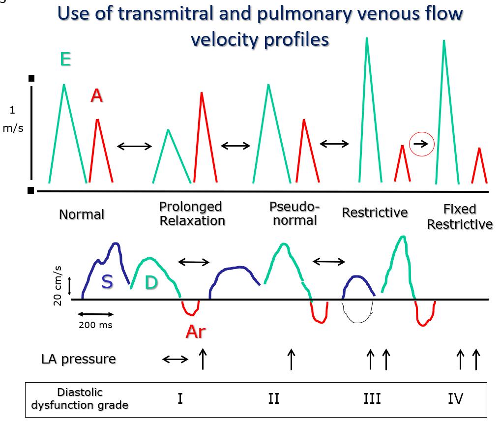
Figure 3. Relationships between transmitral and pulmonary venous flow profiles and correspondence with diastolic dysfunction classes and filling pressures.
The classes of diastolic dysfunction (I to IV) correspond to specific transmitral flow velocity profiles known as “prolonged relaxation”, “pseudo-normal”, “restrictive” and “fixed restrictive”. Class I is characterized by an isolated prolongation of myocardial relaxation. Class II combines an initial reduction in chamber compliance (=increase in stiffness) associated with underlying prolonged relaxation. In class III the reduction of chamber compliance is prevalent over the prolongation of relaxation). Between class I and III there is a progressive increase in filling pressures, which are highest in classes III and IV. Unlike in class III, in class IV the preload reduction maneuvers (nitrate infusion, Valsalva maneuver) are unable to reduce filling pressures. The main advantage of adding pulmonary venous flow profile analysis to mitral valve flow is that it is easier to distinguish the “pseudo-normal” mitral flow profile from normal, because the pulmonary venous profile is “systolic dominant” (prevalence of the systolic wave S) only in normal older subjects and in patients with a prolonged relaxation. In patients with a “pseudo-normal” mitral profile, the pulmonary venous flow profile will show “diastolic dominance” (prevalent diastolic forward D wave). Of note, the peak E wave and deceleration time values of Normal and “Pseudo-normal” profiles are similar. LA: left atrial.
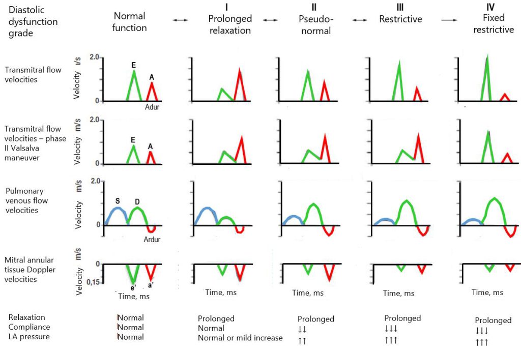
Figure 4. Diagram of the classification of left ventricular diastolic function.
From top to bottom: classes of diastolic dysfunction; transmitral flow velocity profile in baseline conditions and during phase II of the Valsalva maneuver; pulmonary venous flow velocity profile; tissue Doppler velocity profile of the mitral annulus; relationship with left ventricular relaxation, chamber compliance, and left atrial (LA) pressure.
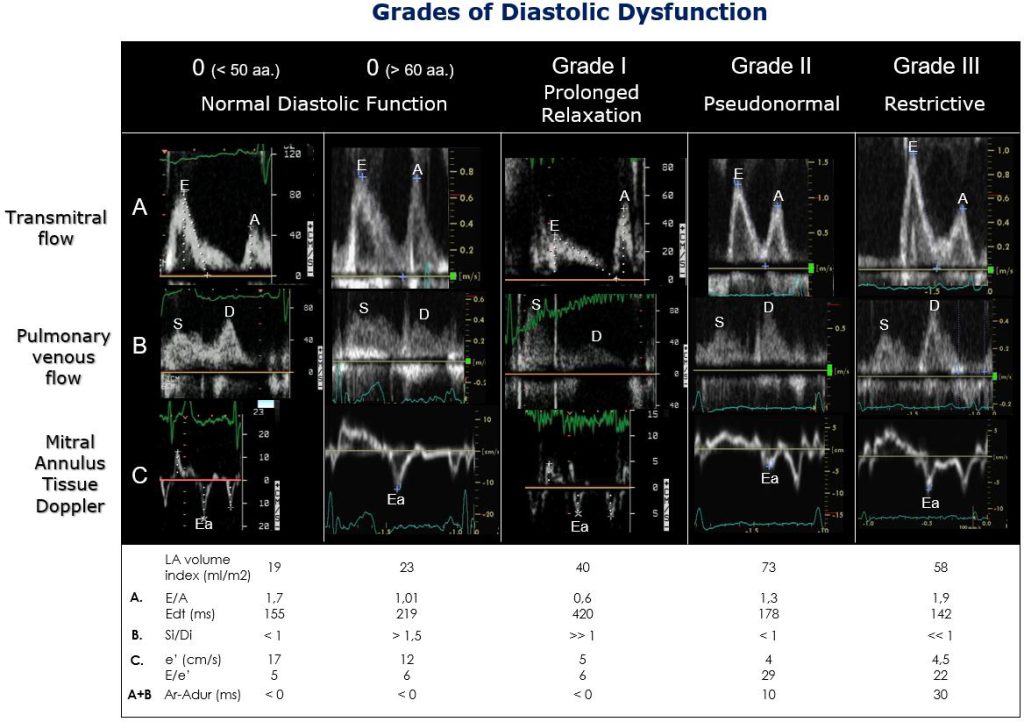
Figure 5. Real-life examples of diastolic dysfunction grades.
Refer to Table 3 for abbreviations and subdivision of normal subjects.
LA: left atrial.