Evaluation of Left Ventricular Stroke Volume
How to calculate ventricular stroke volume using pulsed Doppler
April 24th, 2022
Pages 1 – 5
Definition of Cardiac Output and Stroke Volume
The left ventricular (LV) pump function depends on:
– the contractility of myocardial tissue sarcomeres
– the structure and geometry of the LV chamber
– valvular function
– and the LV loading conditions (preload and afterload)
and can be evaluated at different levels:
– myocardial function (myocardial shortening)
– pump function (ejection fraction)
– cardiac output
We will focus here on the echocardiographic evaluation of Cardiac Output
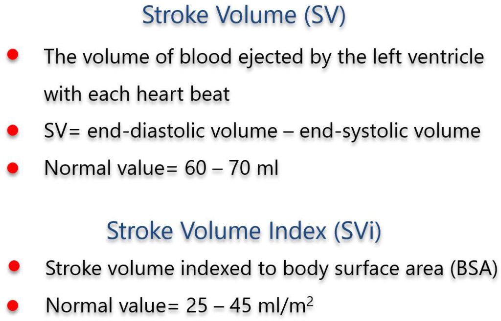
Figure 1. LV stroke volume (SV) is the volume of blood ejected in the main circulation with each single heartbeat. In the absence of valvular regurgitation or ventricular septal defect, the “aortic SV” (the volume of blood passing through the aortic valve in systole) and the “mitral SV” (the volume of blood passing through the mitral valve in diastole) are equivalent. To compare SV between patients, we must calculate the SV index (ml/m2) by indexing to body surface area.
In echocardiography, SV can be estimated with 2D echocardiography (by calculating biplane volumes with the Simpson method), or by using pulsed Doppler of flow through the LV outflow tract or the mitral valve annulus.
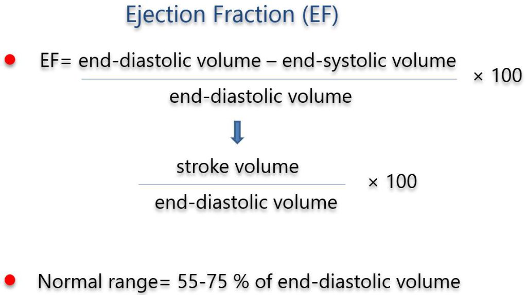
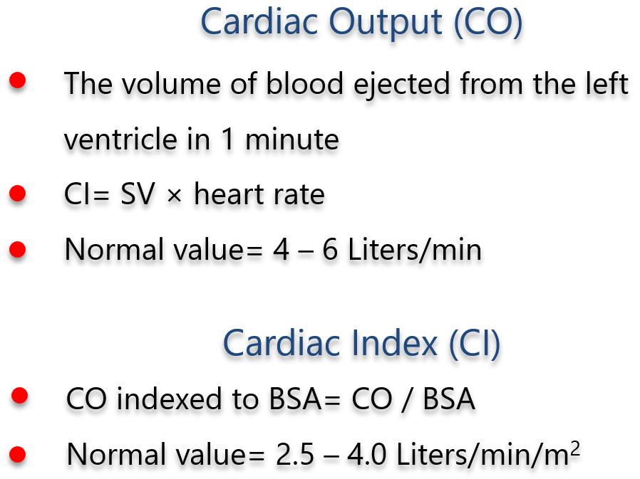
Figure 2. As a matter of fact, LV ejection fraction (EF) is LV SV normalized by end-diastolic volume. As such, LV EF is not a measure of LV contractility, since it is heavily dependent on LV preload, LV afterload and LV intrinsic contractility, and the same holds true for SV and Cardiac Output (Figure 4). The latter is the product of SV by heart rate, and thus measures the volume of blood ejected by the LV in the time frame of 1 minute (Figure 3)

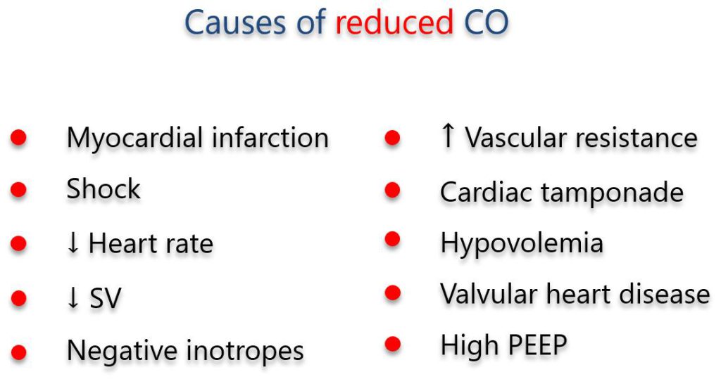
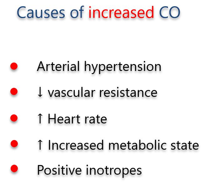
The following are the main causes of a reduction (Figure 5), or an increase (Figure 6) in cardiac output:
PEEP: Positive End Expiratory Pressure (during assisted ventilation).