[Page 2]
Measurement of Flow Volume
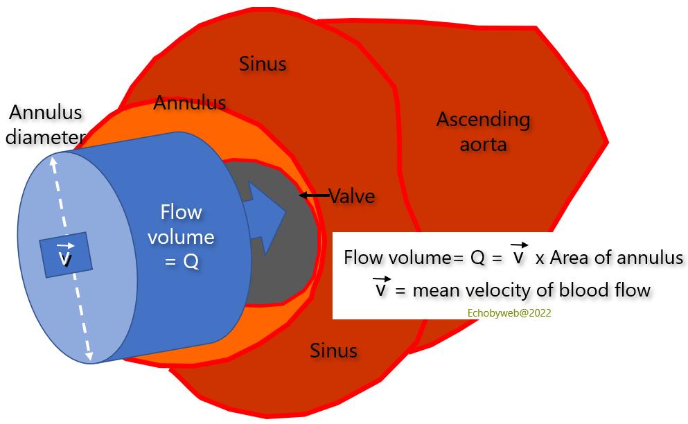
Figure 7. Measurement of Flow Volume.
The volume of flow is the amount of blood crossing the section of a vessel or valve (in this case the aortic valve) in a certain amount of time, for example during systole (aortic or pulmonary valves) or diastole (mitral or tricuspid valves), and it is obtained with the product of the mean velocity of blood flow by the cross-section area of the vessel or valve.
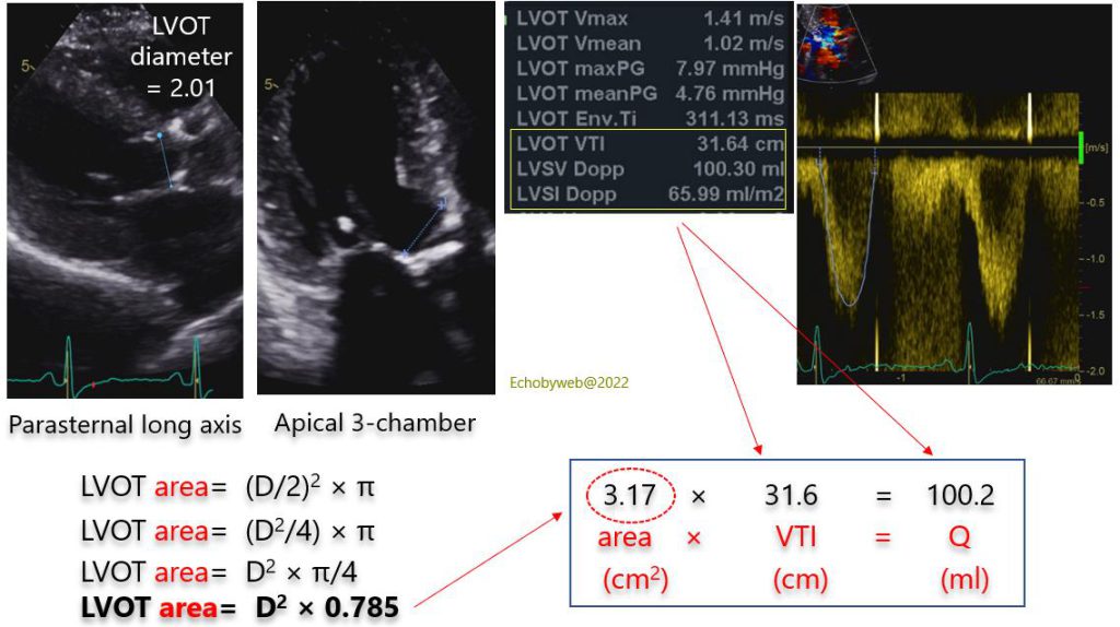
Q= stroke volume, in the absence of valve regurgitation or significant ventricular septal defect. The antero-posterior diameter of the aortic valve annulus is measured immediately below the aortic cusp hinge point, in (preferably) the parasternal long-axis view, or the apical 3-chamber view (Figure 8).
This calculation can be performed also in the right heart, by measuring at the same level the medio-lateral diameter of the pulmonary valve.
The cross-section area of the valve annulus is obtained from the diameter= Diameter2 x 0.785 (Figure 8). The mean velocity of blood flow is obtained by measuring with pulsed Doppler the velocity-time integral of flow (VTI) immediately before the acceleration of flow in the valve annulus (approximately 5 mm proximal to the annulus).
These measurements are obtained at the level of the annulus and not of the tip of the valve cusps (valve area), because it is much easier to calculate the area of the annulus than the area of the valve.
To apply the formula without corrections, it is necessary for the section area of the vessel / valve to not change in time, that is that the flow volume must be only dependent on the variations of the VTI. If the section area changes in time, we must introduce some corrections to calculate the mean area of the valve. Although the aortic annulus is not exactly circular (it is an ellipse), and does modify its area during systole, it is considered adequate to approximate it to a circle with a fixed dimension over time.
The exact positioning of the pulsed Doppler sample volume is very important (there is a learning curve), because it can influence significantly the estimation of stroke volume. For the calculation to be accurate, any cause of blood flow acceleration in the LVOT must be excluded. The evaluation cannot be performed when:
– there is LVOT obstruction (membrane, obstructive hypertrophic cardiomyopathy, severe proximal isolated hypertrophy of the interventricular septum);
– there is a very small LVOT with an acceleration of flow (aliasing velocity set at 50-60 cm/s) which begins before the LVOT;
– there is a mitral valve prosthesis impinging in the LVOT.