Noninvasive Hemodynamics
Noninvasive Hemodynamics: Doppler Measurements of Pulmonary Diastolic Pressure and Left Atrial Pressure
February 14th, 2021
(updated April 14th, 2022)
Pages 1 -2
Table of Contents
- Pulmonary diastolic pressure, continuous wave Doppler … Page 1
- Left atrial pressure, continuous wave Doppler … Page 2
Pulmonary diastolic pressure


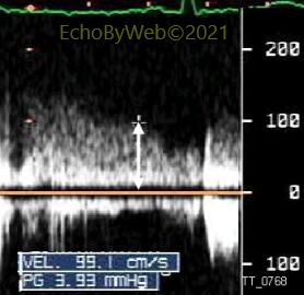
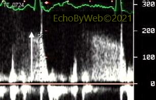
Pulmonary artery diastolic pressure (which approximates pulmonary capillary wedge pressure) = mid-diastolic regurgitant pressure gradient (see below) + RV diastolic pressure (approximated by right atrial pressure, which is estimated from inferior vena cava inspiratory collapse).
Figure 1: Estimated normal wedge pressure. Continuous wave Doppler tracing, transthoracic examination. Normal subject with minimal pulmonary valve regurgitation. End-diastolic gradient= 3.9 mmHg. Assuming normal (= 6 mmHg) right atrial pressure, wedge pressure= 3.9 + 6= 9.9 mmHg.
Figure 2: Estimated increased wedge pressure. Continuous wave Doppler tracing, transthoracic examination. Patient with minimal pulmonary valve regurgitation. Diastolic gradient= 15.2 mmHg. Assuming normal (= 6 mmHg) right atrial pressure, wedge pressure= 15.2 + 6= 21 mmHg.

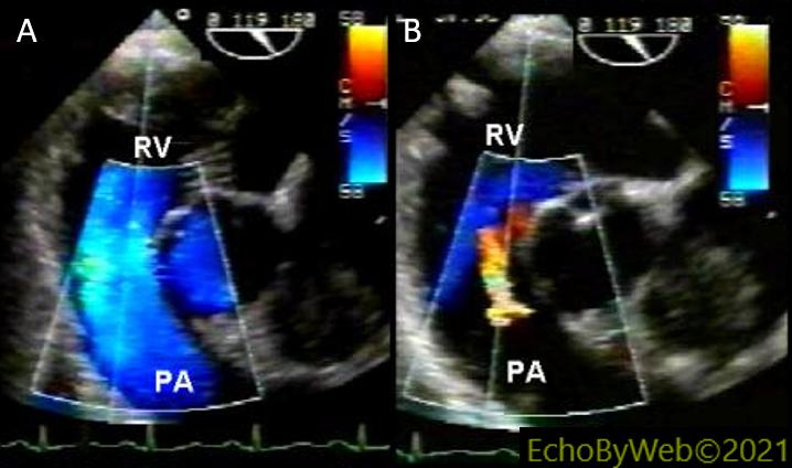
Figure 3. Transesophageal examination, Long axis (119°) of the base, color Doppler flow imaging
A. In blue, normal systolic laminar flow through the right ventricular (RV) outflow tract, valve and pulmonary artery (PA). To the right (and at center of sector scan) short axis of the aortic root.
B: in yellow/red, mild diastolic pulmonary valve regurgitation. The continuous wave Doppler cursor is positioned through, and parallel to, the regurgitant jet, to obtain the diastolic regurgitant flow velocity pattern.