TT Aorta Atheroma Dilatation & Dissection
Transthoracic atlas of the aorta: atheromas, dilatation and dissection
January 30, 2021
(updated April 14th, 2022)
Pages 1 – 2
Table of Contents
- Sino-Tubular Junction: Atheroma 窦管交界处: 动脉粥瘤 … Page 1
- Aortic Arch: Mobile Atheromas 主动脉弓: 可移动的动脉粥瘤 … Page 1
- Aortic Arch Atheroma & Calcification 主动脉弓粥瘤及钙化 … Page 1
- Dilatation of Root & Ascending Aorta 根部扩张及升主动脉 .. Page 2
- Dissection of Ascending Aorta 升主动脉夹层 … Page 2
Sino-Tubular Junction: Atheroma 窦管交界处: 动脉粥瘤
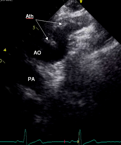
(tt2138) MO, male, 73 y, BSA= 2.03, HR= 87 bpm
HP Sonos 4500 – 2d parasternal long axis – Mild fibrotic thickening of AV cusps.
2d 胸骨旁长轴 – 主动脉瓣瓣叶轻度纤维化增厚。
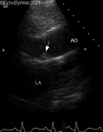
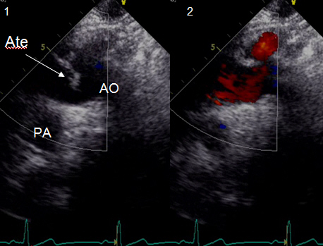
Focused view of aortic root and ascending aorta. A protruding atheroma of posterior sino-tubular junction can be seen in systole (white arrow). The atheroma does not cause flow disturbances (color aliasing) near the posterior aortic wall. Detection of the atheroma must rely on focused imaging of the aortic walls and close inspection of the 2D image
主动脉根部和升主动脉的聚焦视图。 后窦管交界处收缩期可见突出的动脉粥瘤(白色箭头)。 该动脉粥瘤不引起主动脉后壁附近的血流干扰(彩色混淆现象)。 动脉粥瘤的侦测必须依赖于主动脉壁的聚焦视图和对二维图像的仔细检查。
Aortic Arch: Mobile Atheromas 主动脉弓: 可移动的动脉粥瘤
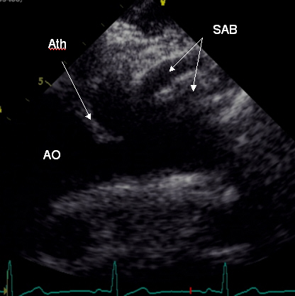
(tt3430) M, 70 y.
Severe mobile atheromas of the aortic arch with thrombi. Pre-operative examination for aneurysm of abdominal aorta.
主动脉弓重度可移动的粥瘤,伴血栓。腹主动脉瘤术前筛查。
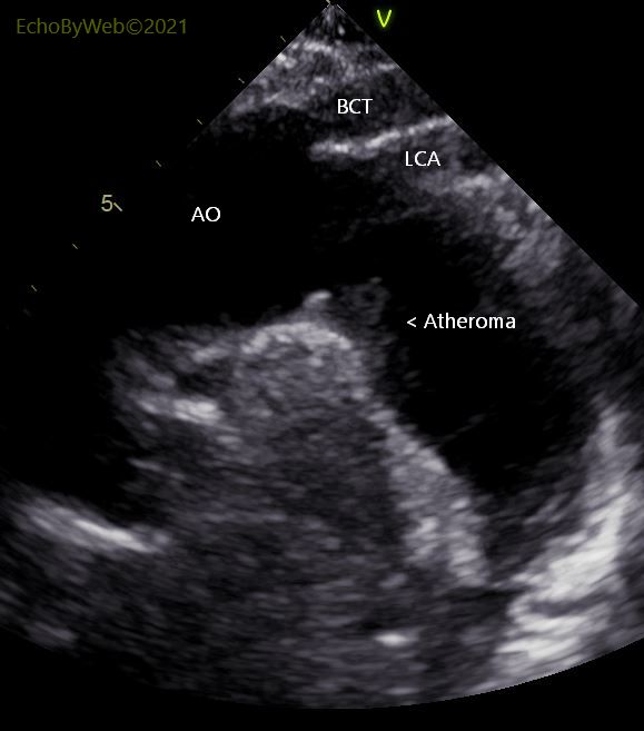
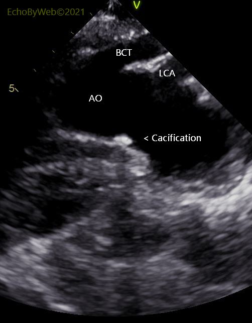
Aortic Arch Atheroma & Calcification 主动脉弓粥瘤及钙化
(#1144) F, 66 y. Ge Vivid95
2D Jugular long axis of aortic arch. 2D 颈动脉长轴主动脉弓
Examination for severe AR. 主动脉瓣重度返流检查
Calcific plaque and atheroma (not mobile) on inner (inferior) wall of the aortic arch.
主动脉弓内(下)壁的钙化斑块和动脉粥瘤(不可移动)。
AO: aortic arch; BCT: brachio cephalic trunk; LCA: left carotid artery.
AO: 主动脉弓; BCT: 头臂动脉干 ; LCA: 左颈动脉