[Page 2]
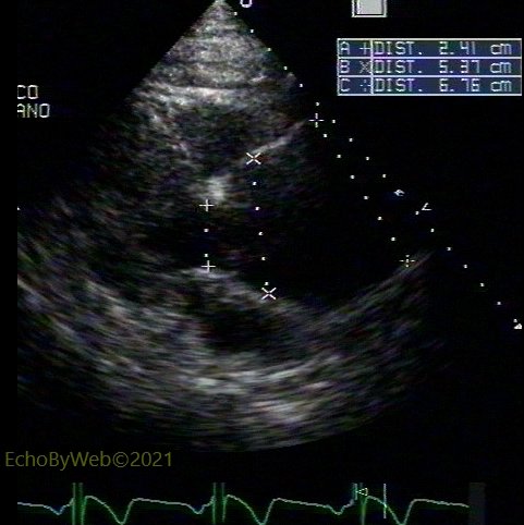
Dilatation of Root & Ascending Aorta 根部扩张及升主动脉
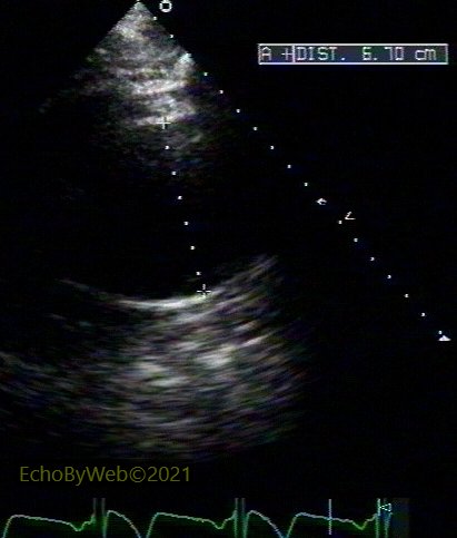
(tt2425) M, 57 y.
Aneurysmatic dilatation of the aortic root and ascending aorta. Mild AR, central.
主动脉根部和升主动脉动脉瘤性扩张。主动脉瓣轻度中心返流。
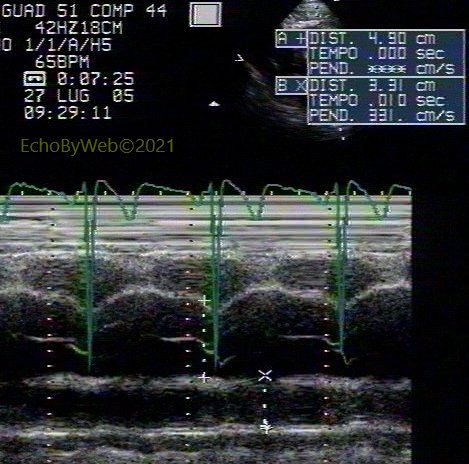
Dissection of Ascending Aorta 升主动脉夹层
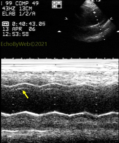
(tt2732) F, 68 y. Sonos 2000. Chest pain and LBBB. Aortic dissection type A, anterior wall of ascending aorta (see intimal mobile flap in 2D parasternal and jugular views, and M-mode) between sino-tubular junction and brachiocephalic branch. Note color Doppler flow in aortic true lumen distal to flap in arch. Associated moderate AR. See also intra-operative TE exam (te0373).
胸痛及左束支传导阻滞(LBBB)。A型主动脉夹层, 升主动脉前壁 (2D胸骨旁和颈动脉视图及M型中可见可移动的内膜皮瓣) 窦管交界处和头臂干之间。 注意主动脉真腔内,即主动脉弓内皮瓣的远端的彩色多普勒血流。符合中度主动脉瓣返流。亦可见于术中食道彩超检查(te0373)
Pages: 1 2