TE Normal 3D Left Atrium
May 15th, 2022
Pages: 1
Normal 3D Anatomy of Left Atrium and Appendage
2D Anatomy
Figure 1. A transesophageal, upper esophagus, 2D short-axis view of the base of the heart (0 deg.). A transverse view of the left atrial appendage is seen to the left (on the right side of the screen) of the aortic root. The left coronary left main trunk up to the bifurcation can be seen between the root and the appendage. The (atrial) systolic contraction of the appendage can be appreciated. Small muscular trabeculae (pectinate muscles) are seen deep in the apex of the appendage. A corresponding orthogonal view (100 deg.) is seen in Figure 2, with the long axis of the appendage which opens in the anterior wall of the atrium. The short axis of the circumflex coronary artery is positioned inferiorly to the base of the appendage.
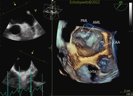
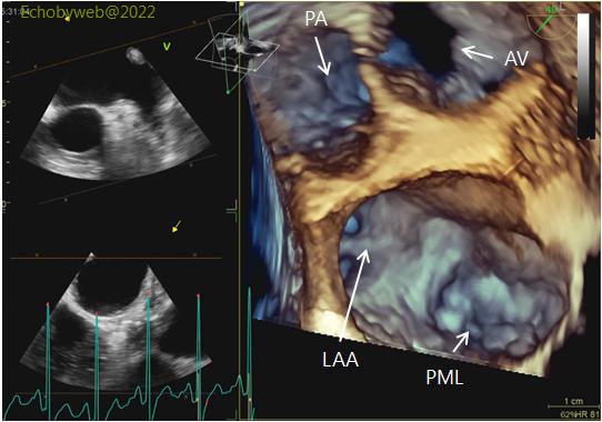
Figure 3. A zoomed real-time 3D reconstruction taken with the TE probe in an intermediate position (49 deg.) between the transverse and longitudinal views. The left atrium is visualized from above (superiorly). Still frames (Figures 4 and 5) show the main anatomical structures. In Figures 3 and 4 the aorta in the image is positioned inferior to the left atrial cavity. In Figure 5, the base of the heart is rotated to obtained the “surgeon’s view” with the aorta superiorly and the left atrium inferiorly in the image, as suggested by the Guidelines.
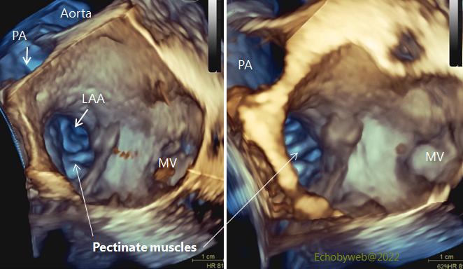
AML and PML: anterior and posterior mitral leaflets; AV: aortic valve; LAA: left atrial appendage; PA: pulmonary artery.
Figure 6. 2 views of the left atrial cavity (surgeon’s view) with slightly different inclinations showing the pectinate muscles inside the appendage cavity. The atrial vestibulum is seen between the appendage and the mitral valve.