Echocardiographic Exam acquisition protocol
An imaging description of the standard echocardiography examination
January 26, 2021
(updated April 29th, 2022)
Pages: 1 – 12
Clinical Echocardiographic Examination Protocol – Brief Operator Guide
General Rules
Please follow the suggested standard viewing sequence, always the same, in every patient you scan: this method increases your accuracy and reproducibility and readability of the exams by your colleagues.
To acquire and maintain a necessary high measurement accuracy, you need to perform all the main measurements in all patients.
It is necessary to save for each view 3 seconds (or 3 heartbeats) video clips, and still images when required (with the performed measurements). The rationale is that it must always be possible to view your measurements on saved examinations, and eventually revise them.
Each exam must have the full patient name
Input patient height and weight. This allows the machine to normalize atrial and ventricular volumes measurements to the BSA (body surface area)
Always connect the ECG cable to obtain a readable ECG trace on the machine.
2D Gain: the blood pool must be black, the pericardium white, the myocardium gray
临床超声检查方针
简要操作指南
总则
请遵循建议的标准图像序列,对每一位患者每次操作都保持一致:这样能提高准确性和高效性,并提高他人可读性。
为了获得并保持必要的测量高准确度,要在每位患者做检查时获取全部的测量
保存每个三秒长的动态图像是很有必要的(或3个节律),需要时还要获取静态图像(包含测量)。这是为了保证随时能在已存的超声检查中看到你获取的测量,及最终对其修正。
每个检查必须写有患者姓名
输入患者身高体重。这样能使机器根据体表面积规范出正常的心房心室容积
每次都要连心电,使在机器上能获取准确的心电轨迹
2D增益值:血池颜色必须是黑色,心包白色,心肌灰色
Acquisition sequence for each view
· 2D imaging video clip (3 heart beats)
· 2D still frame with measurements (if required)
· Color Doppler video clip
· M-mode still frame with measurements, if required
· Pulsed Doppler still frame with measurements, if required
· Continuous wave Doppler still frame with measurements, if required
· Dopper Tissue Imaging still frame with measurements, if required
每个视图的图像获取序列
· 2D动态图像(3个节律)
· 2D图像含测量(如果需要)
· 颜色多普勒动图图像
· M模图像含测量,如果需要
· 脉冲波多普勒图像含测量,如果需要
· 连续波多普勒图像含测量,如果需要
· 组织多普勒图像含测量,如果需要
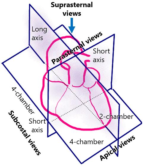
Figure 1. The image shows the available echocardiographic transthoracic views.
It is evident that:
– The long axis views of the left and right ventricles from the parasternal and apical (3-chamber) views are equivalent
– The short axis of the left and right ventricles from the parasternal and subcostal views are equivalent
– The 4-chamber views as explored from the apical or subcostal windows are equivalent
Thus, the parasternal imaging in a patient with poor anterior thorax ultrasound penetration (ex: emphysema) can be successfully substituted by apical and especially subcostal imaging.
Imaging Protocol