[Page 4]
B. Parasternal Long Axis, Right Heart – 胸骨旁长轴,右心

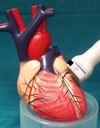
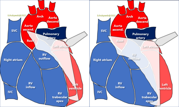
2D video clip. Right ventricular long axis, obtained by tilting the transducer medially (to the right of the patient), starting from the parasternal long axis of the left ventricle, and “crossing” the interventricular (anterior) septum (Figure 14).
Position markers, visualization of:
1. maximum diastolic excursion of tricuspid leaflets;
2. Inferior vena cava;
3. Maximum right ventricular longitudinal axis.
This view evaluates the right ventricle, tricuspid valve, right atrium, coronary sinus. It is the only view that visualizes the anterior right ventricular free wall.
Exclude pericardial anterior effusion / adhesions.
2D动态图像. 右室长轴(右室、右房、三尖瓣、冠脉窦

CD video clip. Tricuspid valve flow: locate and maximise regurgitant jet location / direction, if any. Exclude aliasing secondary to stenosis. Evaluate flow from inferior vena cava and coronary sinus with color Doppler and pulsed Doppler if necessary.
彩色多普勒动态图像. 三尖瓣血流;放大图像,找到返流的射流位置/方向

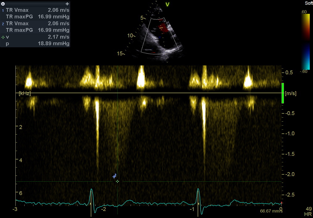
Continuous Wave Doppler still frame. Tricuspid regurgitation: measurement of peak mid-systolic velocity. Purpose: to estimate right ventricular systolic pressure (see online Calculator here), combined with estimation of right atrial pressure (inspiratory collapsibility of the inferior vena cava, see Page 12).
This measurement – together with the right ventricular outflow tract time-velocity integral – is currently used in a validated formula to estimate also pulmonary vascular resistances (see Online Calculator here).
Note that peak tricuspid regurgitant velocity should be evaluated in multiple views.
连续波多普勒图像. 三尖瓣返流, 测量:最大流速