[Page 12]
H. Subcostal views – 腱突下

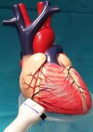
2D video clip. Subcostal 4-chamber view. Position markers, visualization of:
1. Maximum left ventricular longitudinal axis;
2. Maximum diastolic excursion of mitral valve leaflets.
Evaluate the right ventricle (optimize chamber area) and left ventricular dimensions and wall motion.
When the parasternal views are not available, left ventricular linear and volume (monoplane 4-chamber) measurements can be performed here.
2D动态图像. 4腔图,评估右室(优化右室区域),评估左室尺寸和室壁运动
CD video clip. Subcostal 4-chamber view centered on the interatrial septum. Evaluate septal integrity.
Evaluate flow from hepatic veins.
彩色多普勒动态图像. 4腔图,房间隔放中间;评估间隔完整性

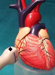
2D video clip. Subcostal bi-caval. Optimise view initially for the inferior vena cava, and then for superior vena cava. Position markers, visualization of:
1. maximum expiratory diameter of inferior vena cava;
2. maximum diameter of inferior vena cava at entry into the right atrium.
Pulsed Doppler analysis of vena cava flow velocities, and respiratory variability.
Evaluate inspiratory collapsibility. This measurement, combined with the measurement of peak systolic tricuspid regurgitant velocity (see above) is used to estimate right ventricular systolic pressure (see Online Calculator here)
2D动态图像. 下腔静脉优化图像; 评估呼吸塌陷