[Page 2]
Imaging Protocol
A. Parasternal Long Axis, Left Heart – 胸骨旁长轴,左心

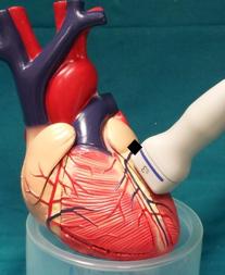
2D video clip. Parasternal left ventricular long axis, Position markers (to identify correct positioning of scan sector), visualization of:
1. Maximum diastolic excursion of mitral leaflets;
2. Maximum systolic excursion of aortic cusps;
3. Maximum LV longitudinal axis;
4. Short axis of coronary sinus.
This view is very important, it gives an immediate overview of the heart’s global functional status. The structures that can be analyzed, from left to right (check anatomy and function of) are: aortic valve, left atrium, coronary sinus, thoracic descending aorta, mitral valve, right ventricular dimensions and anterior wall thickness, left ventricular outflow tract, left ventricular dimensions and wall thickness of the interventricular septum and infero-posterior wall; evaluation of left ventricular wall motion. Exclude anterior and posterior pericardial effusion / adhesions.
In a left ventricle with normal dimensions and stroke volume, the anterior mitral leaflet in early diastole should almost touch the anterior proximal septum (within 1 cm ) (Figure 6), the anterior septum and infero-posterior walls thicken homogeneously and move towards the center of the cavity. The aortic cusps in systole open completely, coming in close reach of the aortic root sinuses (Figure 5). The atrial cavity expands in systole almost 100%, indicating a normal stroke volume. The right anterior wall contracts and thickens, and anterior to the wall the epicardium slides underneath the pericardium, indicating normal pericardial function.
2D 动态图像 左室长轴(主动脉瓣、左房、二尖瓣、左室、室间隔、后侧壁、降主动脉);评估室壁运动

2d still frame 2D. Left ventricular measurements.
End-diastole: interventricular septal thickness, left ventricular diameter, infero-posterior wall thickness; measurements should be performed at the level of the tips of the mitral leaflets; mitral E-septal separation.
Mid-systole: left ventricular outflow tract diameter, which is measured immediately below the hinge points of the cusps.
See Normal values here.
图像
左室的测量: 舒张末期:室间隔厚度,左室直径,后侧壁厚度. 收缩中期:左室流出道直径
End-systole: left ventricular end-systolic diameter. If M-mode is used, the exact time point is at the notch between the maximum systolic thickening and the (physiologic) early diastolic rapid posterior movement of the anterior septum.
2D linear measurements are preferable to M-mode measurements because in the former case it is possible to obtain measurements perpendicular to the ventricular long axis, and avoid overestimation of septal thickness in the frequent event of localized proximal anterior septal hypertrophy (which has no clinical consequences and is not a marker of left ventricular hypertrophy.