[Page 7]
E. Apical Views, Evaluation of the Left Atrium – 心尖图
2D video clip. Apical 4-chamber view, optimized for left atrial visualization (priority should be given to obtaining a maximal left atrial long axis).
Position markers, visualization of:
1. Maximum left atrial longitudinal axis;
2. Maximum left atrial transverse axis;
3. Maximum diastolic excursion of the mitral leaflets
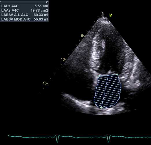
2D still frame. 4-chamber view, centered on the left atrium. Measurement: left atrial planimetry to obtain the biplane volume (see below the 2-chamber view). See Normal values here.
2D图像. 4腔图,左房放中间,测量:双平面侧面法测左房容积
2D video clip. 2-chamber view, optimized for left atrial visualization (priority for left atrial long axis visualization).
Position markers, visualization of:
1. Maximum left atrial longitudinal axis;
2. Maximum diastolic excursion of the mitral leaflets
3. Short axis of coronary sinus (inferior);
4. Left atrial appendage (anterior).
2D动态图像. 2腔图,左房放中间
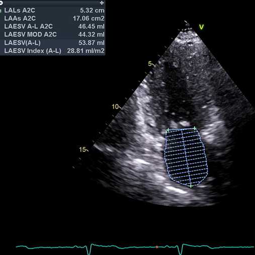
2D still frame. 2-chamber view, optimized for the left atrium. Measurement: left atrial planimetry for biplane volume (see also 4-chamber view above). See Normal values here.
2D图像. 2腔图,优化左房图像,测量:双平面侧面法测左房容积