LV Analysis, Morphology
Measurement of Left Ventricular 2D Mass Index
February 14th, 2021
(update April 14th, 2022)
Left Ventricular Mass Index, 2d
LV mass (area/length). 2D (gm)
1.05 x ([5/6 x A1 x (a + d + t)] – [5/6 x A2 x (a + d)]) [Helak, 1981]
LV mass (truncated ellipsoid), 2D (gm):
1.05 p {(b + t)2 x [2/3 x (a + t) + d – [d3 / 3 x (a + t)2]] – b2 x [2/3 a + d – (d3 / 3 x a2)]} [Schiller, 1989]
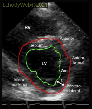
Figure 1 Transthoracic parasternal short axis view. Red: tracing of pericardial border to obtain A1. Green: tracing of endocardial border (papillary muscles are excluded) to obtain A2. Am = A1 – A2 = area of myocardium
t: myocardial thickness (automatically calculated by the software).
LV mass index (truncated ellipsoid) normal values:
Males: 76±13 gm/m2
Females: 66±11 gm/m2
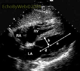
Figure 2. Transthoracic subcostal 4-chamber view .a: semi-major axis from widest minor axis radius (b) to apex; b: short axis radius; d:truncated semi-major axis from widest minor axis radius (b) to mitral anulus plane.
LA: left atrium; RA: right atrium; RV: right ventricle