TE Bicuspid Aortic Valve & Mild Stenosis
May 30, 2022
Pages 1
See transthoracic case presentation here (Transthoracic Atlas)
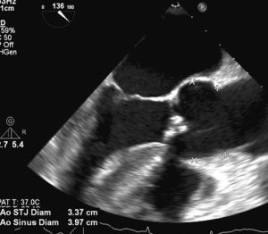
Follow-up of transthoracic case presentation. The aortic valve is bicuspid (Figures 2-4) with a prevalent antero-lateral cusp and a postero-medial cusp. There are nodular calcifications and no signs of significant stenosis at continuous wave Doppler sampling.
Because of the moderate dilatation of the ascending aorta (50 mm), the patient underwent aortic valve replacement with a bileaflet mechanical prosthesis and replacement of ascending aorta. Recovery was uneventful.