Analysis of Aorta
Echocardiographic Measurements of the Aorta
February 14th, 2022
(updated April 29th, 2022)
Pages 1 -2
Table of Contents
- Aortic root diameter, M–mode
- Aortic root & sino-tubular junction, 2D
- Left ventricular outflow tract / aortic valve annulus diameter, 2D
- Aortic valve diameter for the implant of a percutaneous / transapical biologic aortic valve, 2D
- Thoracic ascending aorta
- Aortic arch, 2D
- Aortic arch, color Doppler
- Descending thoracic aorta, 2D
- Ascending aorta and arch atheromas
Aortic root diameter, M-mode
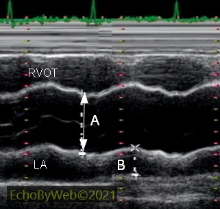
In M-mode, parasternal long axis view, the antero-posterior diameter of the aortic root is measured with the “leading-edge to leading-edge” method (A), synchronous with the Q wave of the EKG.
B: antero-posterior diameter of the left atrium (LA), measured at maximum atrial expansion in end-systole.
LA: left atrium; RVOT: right ventricular outflow tract.
In the example, a moderate to severe dilatation of the aortic root (54 mm).
Aortic root and sino-tubular junction diameters, 2D
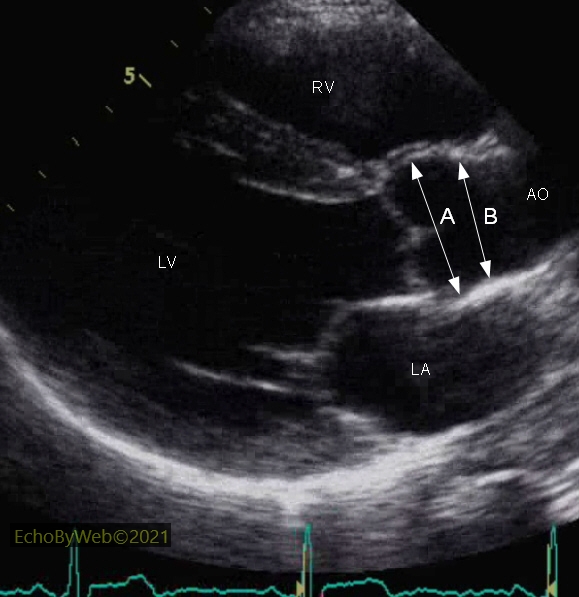
In 2D mode, parasternal long axis view, the maximum antero-posterior aortic root (A, between the anterior right coronary and posterior non coronary sinus of Valsalva) and the minor sino-tubular junction (B) diameters are measured between the internal borders (intima) of the aortic walls, in end-diastole (ECG Q-wave).
LA: left atrium; LV: left ventricle; RV: right ventricle, outflow chamber.
The 2D measurement of the aortic root is smaller than the corresponding M-mode measurement (see: figure A), but it can be compared to the corresponding CAT scan measurement.
In the example, a normal exam.
Left ventricular outflow tract / aortic valve annulus diameter, 2D
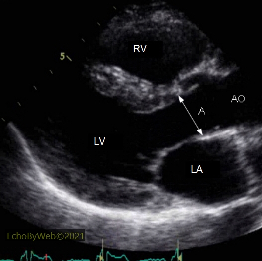
In 2D mode, parasternal long axis view, the outflow tract / aortic valve annulus diameter (A) is measured in systole between the outflow tract anterior and posterior endocardiums immediately below the attachments of the anterior (right coronary) and posterior (non coronary) aortic valve leaflets.
AO: aortic root; LA: left atrium; LV: left ventricle; RV: right ventricle.
This measurement is used to calculate with the continuity equation the functional area of the native aortic valve (aortic stenosis) or of the aortic valve prosthesis.
Aortic valve diameter for the implant of a percutaneous / transapical biologic aortic valve, 2D
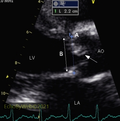
In 2D mode, parasternal long axis view, the annulus (A) is measured between the anterior and posterior most external borders of the native calcific (stenotic) aortic valve , in systole. For comparison purposes, the measurement of the standard LV outflow / aortic annulus (B) used in the calculation of the continuity equation (see also figure C).
AO: aortic root; LA: left atrium; LV: left ventricle.
The measure is quite demanding, and requires the differential visualization of the anterior aortic root wall and the calcific aortic valve tissue. (the arrow points at the calcific anterior right coronary aortic valve cusp).
Thoracic ascending aorta

In 2D mode, parasternal long axis view, the maximum antero-posterior diameter of the ascending thoracic aorta is measured between the inner borders (intima) of the walls (A). Although the Guidelines recommend to perform the measurement using the leading edge to leading edge technique, I personally suggest to perform the measurement as indicated, because it logically corresponds to the same measurement performed with a CT scan.
The measurement should be performed where the maximum diameter is visualized, noting preferably also the distance between this measurement and the aortic valve plane (B).
The ascending aorta is frequently visualized 1-2 intercostal space superior to the one utilized for the parasternal long axis view of the left ventricle. It is not necessary to visualize in the same view of the ascending aorta also the left ventricular and atrial cavities.