TE Neocuspidization Procedure
Transesophageal Atlas of Valve Prosthesis: Ozaki procedure
February 13th, 2022
(updated April 14th, 2022)
Pages 1 – 3
Aortic Valve Neocuspidization Intervention (Ozaki)
Figure 1. TE exam pre-CPB. Severe aortic regurgitation, with central origin, as seen from the transgastric 5-chamber view
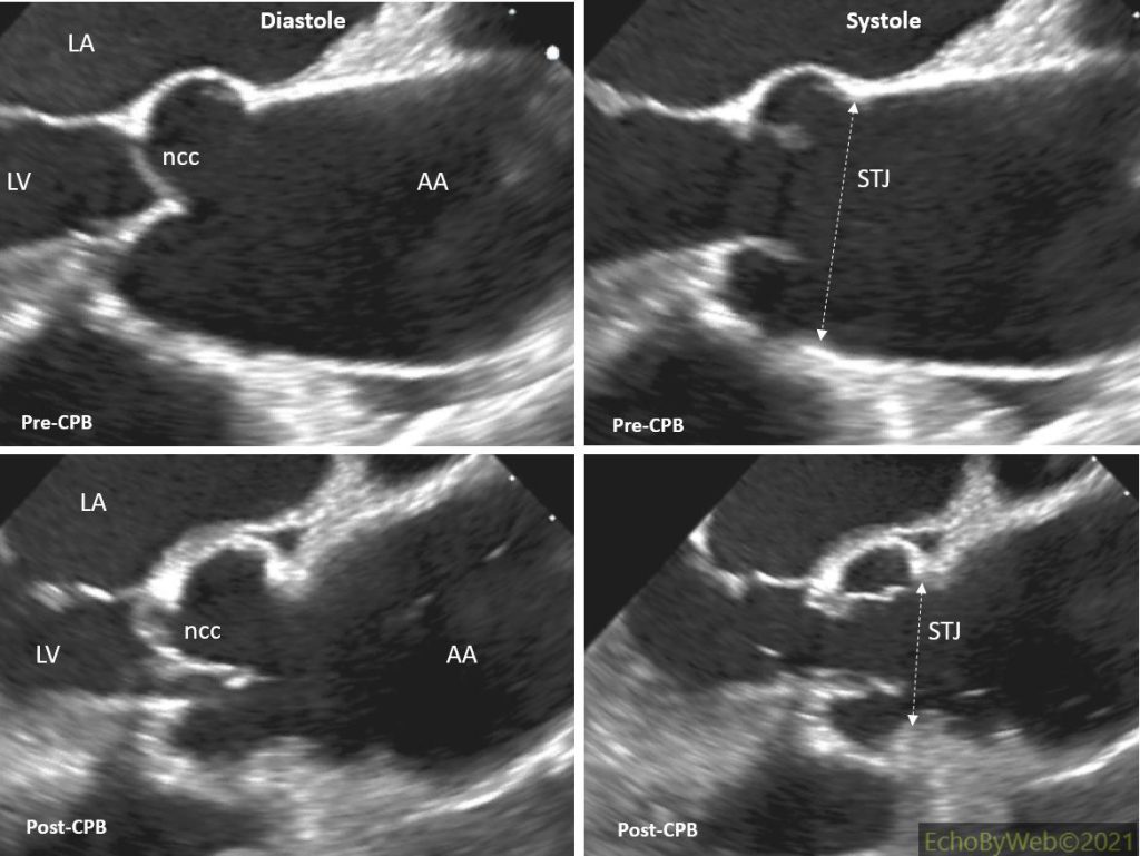
Figures 2-4. TE exam pre- and post-CPB. In the post-CPB images the AV neo-cusps are clearly seen with opening and closure dynamics similar to those of the native cusps. Notice the long apposition line in diastole which reaches the SDJ (sino-tubular junction) plane (dotted arrows).
AA: ascending aorta; LA: left atrium; LV: left ventricle; ncc: posterior non-coronary cusp.