[Page 4]
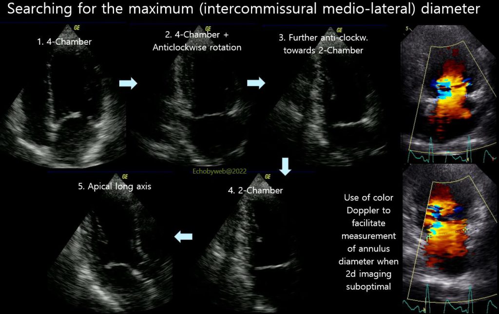
Figure 11 shows how to search for the maximum inter-commissural diameter. Starting from the apical 4-chamber view, the maximum mitral annulus diameter is found in the intermediate views between the 4-chamber and the 2-chamber views (Figure 11, 1 to 4), with careful anticlockwise rotation of the transducer. The transducer may be moved to an upper intercostal space (compared to that used for the 4-chamber view) during rotation. It is important to maintain a central position for the LV long axis.
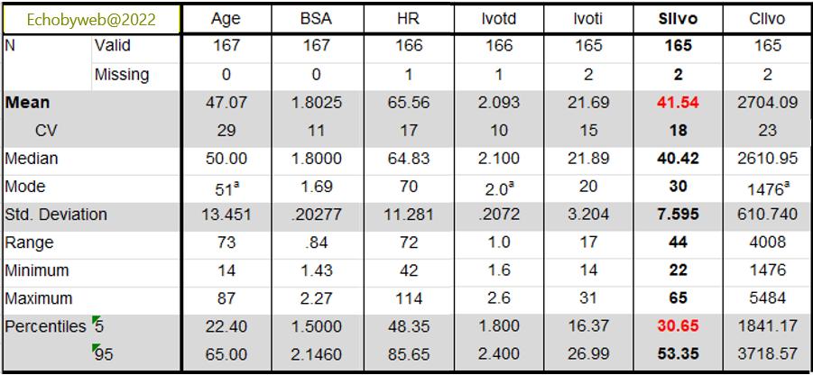
Normal values for pulsed Doppler derived Stroke Volume index and Cardiac index from a group of 165 normal subjects are shown in Figure 12 (personal data of Author).
HR: heart rate; lvotd: LV outflow tract diameter; lvoti: lV outflow tract pulsed Doppler flow VTI: SIlvo: stroke index; CIlvo: cardiac index.