[Page 6]
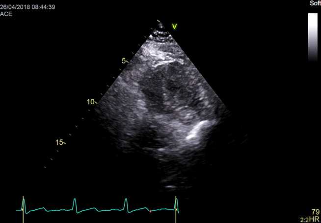
D. Parasternal Short Axis, Mitral Valve – 胸骨旁短轴
2D video clip. Short axis of the left ventricle, at the level of the mitral valve. Position markers:
1. With caudal scanning, visualize the continuity between the anterior mitral leaflet and the posterior noncoronary aortic cusp;
2. Identify the correct anatomy of the antero-lateral and postero-medial commissures.
Evaluation of mitral valve leaflet dynamics.
2D动态图像. 左室底部短轴,二尖瓣放中间;评估室壁运动
CD video clip. Mitral valve flow. Locate the position and dimensions of the Regurgitant Orifice Area of regurgitation.
彩色多普勒动态图像. 二尖瓣血流;找到有效返流口
D. Parasternal Short Axis, Left Ventricle – 胸骨旁短轴

2D video clip 2D. Short axis of the left ventricle, papillary level.
Position markers, visualization of:
1. Cranial scanning: continuity between papillary muscles, chordae and MV commissures;
2. Postero-medial and antero-lateral papillary muscles.
Evaluation of wall motion: see here for grading of wall motion.
动态图像. 左室短轴,乳头肌水平;评估室壁运动
2D video clip. Short axis of the left ventricle, apical level; evaluation of wall motion
2D动态图像. 左室短轴,心尖水平;评估室壁运动
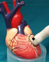
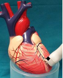
Figure 34 and 35. Transthoracic parasternal short-axis view. Differences between scanning by tilting the transducer (without displacing it, left panel) or by translating the transducer towards the ventricular apex (right panel). Although the tilting methos can be used to obtain an initial fast assessment of left ventricular anatomy, analysis of left ventricular wall motion or any specific evaluation (ex: evaluation of mitral regurgitant orifice area) should be performed by translating the transducer on the thorax, so to keep the scan sector perpendicular to the left ventricular long axis.