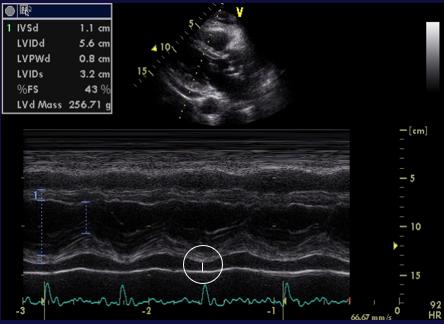
Figure 28 (tt2957). M-mode parasternal long axis, left ventricle
Above: Figures 26-28. Mild to moderate pericardial effusion (11 mm). 2D (Figure 27) and M-mode (Figure 28) quantification.
Below: Figures 29-34. Severe diffuse effusion without tamponade. There is collapse of the RA and RV walls (Figure 33) without paradoxical pulse. Note the “swinging heart” sign that can be observed from all views. Figure 34 shows a dilated inferior vena cava with preserved inspiratory collapse (= normal estimated RA pressure).
Figure 29 (tt2435). 2D parasternal long axis
Figure 30 (tt2435). 2D parasternal short axis
Figure 31 (tt2435). 2D parasternal short axis scan
Figure 32. 2D measurements of the severe pericardial effusion shown in Figures 29-31
We use technologies like cookies to store and/or access device information. We do this to improve browsing experience and to show (non-) personalized ads. Consenting to these technologies will allow us to process data such as browsing behavior or unique IDs on this site. Not consenting or withdrawing consent, may adversely affect certain features and functions.
Functional
Always active
The technical storage or access is strictly necessary for the legitimate purpose of enabling the use of a specific service explicitly requested by the subscriber or user, or for the sole purpose of carrying out the transmission of a communication over an electronic communications network.
Preferences
The technical storage or access is necessary for the legitimate purpose of storing preferences that are not requested by the subscriber or user.
Statistics
The technical storage or access that is used exclusively for statistical purposes.The technical storage or access that is used exclusively for anonymous statistical purposes. Without a subpoena, voluntary compliance on the part of your Internet Service Provider, or additional records from a third party, information stored or retrieved for this purpose alone cannot usually be used to identify you.
Marketing
The technical storage or access is required to create user profiles to send advertising, or to track the user on a website or across several websites for similar marketing purposes.