[Page 14]
Classification of constrictive pericarditis.
- Typical
– Chronic
– Subacute - Effusive-constrictive
- Localized
- Occult
- Transient
Transient constrictive pericarditis.
In some patients with acute constrictive pericarditis, the symptoms and constrictive physiologic features resolve with medical therapy alone. The causes of pericarditis are diverse, the most common prior cardiovascular surgery.
Haley JH. Transient constrictive pericarditis. Causes and natural history. J Am Coll Cardiol 2004; 43:271.
Effusive-constrictive pericarditis.
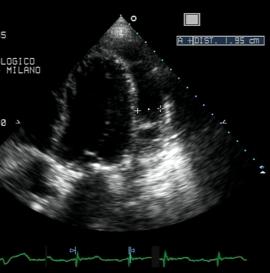
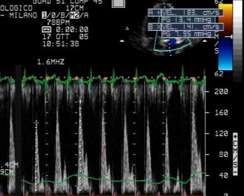
Figures 91-93. Male, 45 year old patient, status postoperative for aortic mechanical valve prosthesis, mitral and tricuspid valve repairs. Post-pericardiocentesis exam. EDV= 90 ml/m2; EF= 55%; LA volume index 40 ml/m2; pulmonary systolic pressure= 37 mmHg. There is residual moderate effusion (Fig. 92) lateral to the LV with fibrotic organization. 2D analyiss shows typical leftwards septal shift with inspiration (Fig. 91) and pulsed Doppler analysis of mitral valve flow shows peaking of E wave at first expiratory beat (Fig. 93). These findings are consistent with pericardial constriction.
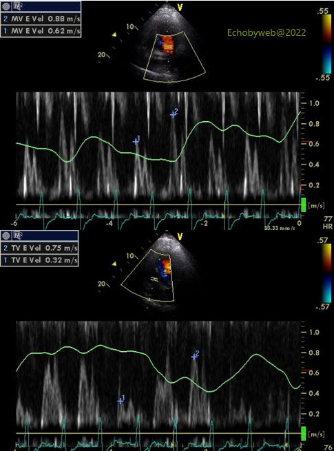
Figures 94-96. Status post mitral valve replacement with bioprosthesis. At 2D analysis there are both adhesions (apical Fig. 94) and organized effusion (Fig. 95). Pulsed Doppler echocardiography shows typical signs of constriction (41% increase in peak mitral E wave and 134% increase in peak tricuspid E wave).
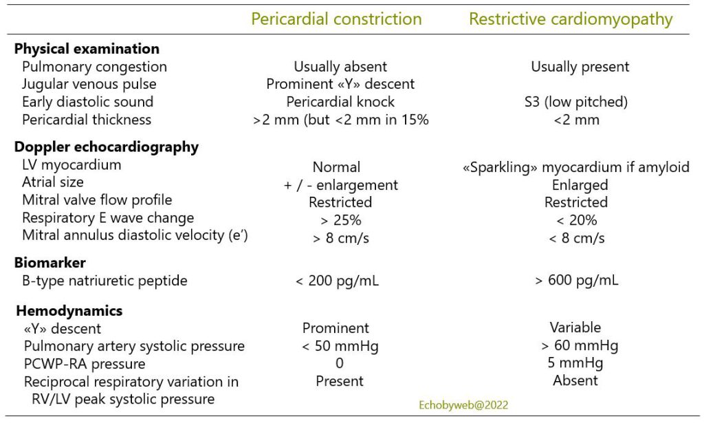
Differential diagnosis between pericardial constriction and restrictive cardiomyopathy.
Prognosis.
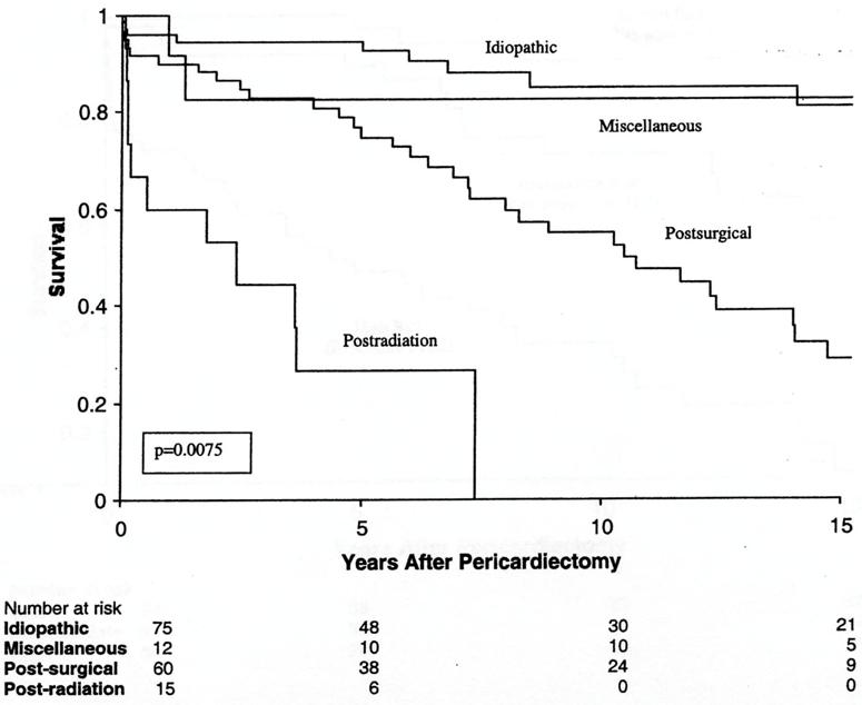
Figure 97. Bertog et al. Constrictive pericarditis: etiology and cause-specific survival after pericardiectomy. J Am Coll Cardiol 2004;43:1445 – Cleveland Clinic