[Page 7]
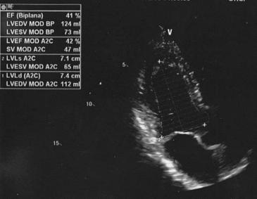
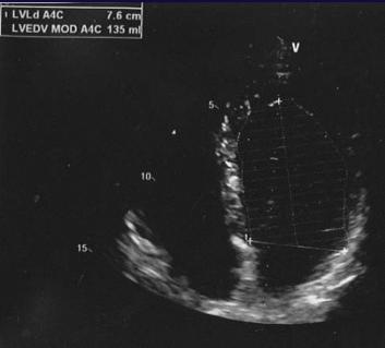
This outpatient exam (Figures 22 -23), performed in October 2013, reports mild concentric hypertrophy, mild mitral regurgitation, global LV hypokinesis and EF= 39 %. The operator suggests in the conclusions to admit the patient for “coronary arteriography and ventriculography”, which the patient refuses. As discussed previously (Figures 1-8, Page 2), the 4-chamber view is clearly foreshortened with a circular area.
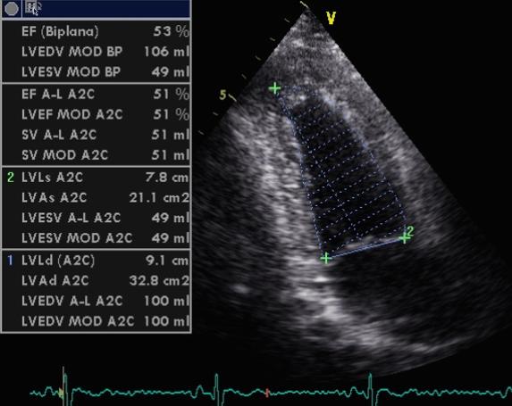
This second exam (Figure 24-25), requested by the patient 2 days later at another laboratory, shows an EF at lower normal limits= 53 %, and normal segmental wall motion. Compare the area of the cardiac fossa with that shown in Figure 23.
The patient complained of what appeared to be atypical chest pain.