[Page 5]
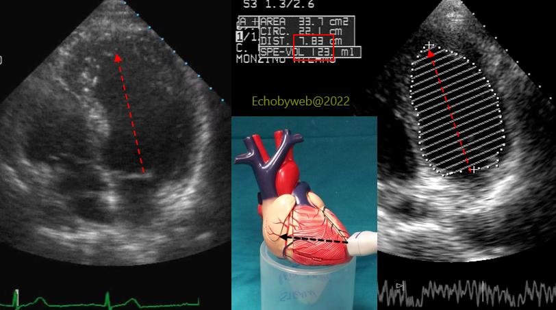
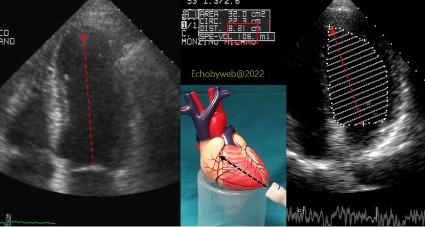
Another example: above (Figure 14) incorrect transducer position on the apical anterior wall, foreshortening of the LV long axis (7.8 cm) and overestimation of the LV end-diastolic volume (123 ml). Below (Figure 15), correct positioning of the transducer on the true LV apex: the correct LV long axis= 8.2 cm, and end-diastolic volume= 106 ml.
This is a 43 year old patient with previous AMI and CABG.
In this patient, also the 2-chamber has been acquired incorrectly (Figure 16). Compare to the correct visualization of the apical 2-chamber (Figure 17): the long axis is foreshortened and the RV cavity can be seen inferiorly to the inferior apex (Figure 16), pointing to a misalignment of the view. The plane of the image is not through the midplane of the true 2-chamber view, but is displaced medially. This causes a false normokinesis / hyperkinesis of the LV apical segments.