[Page 7]
Information provided by the echocardiographic examination.
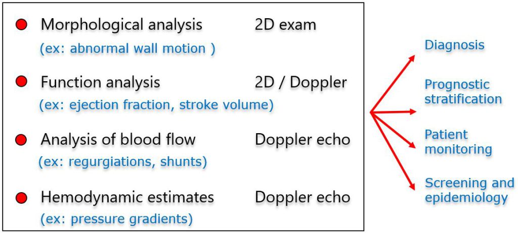
The “4 layers” of information provided by the exam. The operator should approach echocardiographic imaging keeping in mind the progressive acquisition of all these information categories.
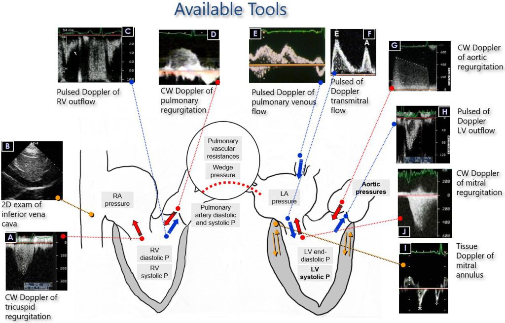
Figures 27 and 28. An example of the available “tools” that can be used to acquire the 4th layer of information: Hemodynamic estimates (see for details: “Classroom -> LV Diastolic Function“, Page 8).
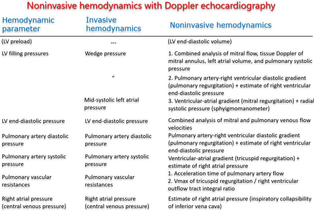
Figure 28. Noninvasive 2D, pulsed and continuous wave Doppler measurements of hemodynamic parameters.