[Page 4]
Image acquisition: technical considerations.
- Patient / operator positioning
- ECG trace
- Optimization of LV area
- Respiration control
- Digital acquisition and recording of images
- Side by side comparison with previous studies
- On-line measurements
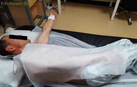
Patient position:
- On his left side (if operator on the right of patient)
- Pillow for comfort / head rest
- Clean paper sheet on pillow and mattress
- Paper sheet between you and patient
- Left arm on patient side: patient more comfortable
- Right arm stretched out or under head: you can reach with your tranducer high near armpit when LV long axis is horizontal
- Better if patient undressed: it is easier to reach transducer positions
This listing may seem obvious, but in some workplaces it still needs to be implemented
Influence of patient position on imaging quality.
Figures 8 and 9. On the left, imaging obtained in a patient in a semi-left recumbent position: incomplete / non-optimal visualization of the endocardium of the LV lateral wall. On the right, patient in a complete left lateral recumbent position: optimal visualization of the LV endocardium.
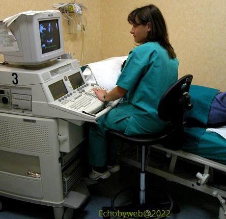
Operator ergonomics.
- Make use of the possibility to adjust all available equipments to obtain an adequate upright posture with the arms as close as possible to your body, and your head aligned with the torso, and both upright.
- This may require to adjust the height of the examination bed / your chair to reduce the distance from patient. Move the patient close to the edge of the bed (towards you).
- In particular, pay attention to the posture of your neck, shoulders and back.