[Page 3]
Figure 9 shows the 3D rendering of the mitral valve (right panel), as viewed from the left ventricle. It is possible to appreciate the fusion of the postero-medial commissure (on the left), whereas the antero-lateral commissure (on the right) is not clearly seen (echo dropout). Calcification and fusion of the chordae from the posterior papillary muscle is seen above the posterior commissure.
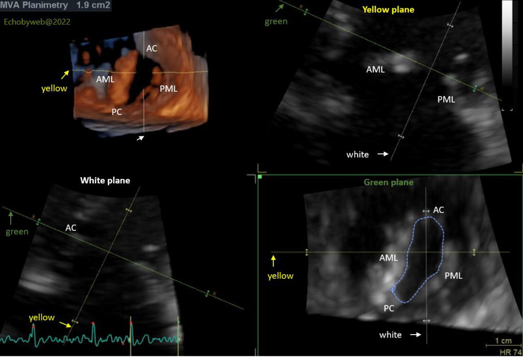
Figure 10 shows the 3D reconstruction of the mitral valve orifice with the GE FlexiSlice instrument. The mitral valve area at planimetry is 1.9 cm2, which corresponds to the 2D planimetry (Figure 3), and is greater than the area derived from the PHT (Figures 4-6).
Upper left panel: 3D rendering of the mitral valve as seen from the left ventricle. Lower right panel (the Green plane of the 3D data): short axis plane of the mitral valve, as selected by positioning the green plane lines in the upper right (yellow antero-posterior plane) and lower left (white intercommissural plane) panels.
AC: anterior commissure; AML: anterior mitral leaflet; PC: posterior commissure; PML: posterior mitral leaflet.
This 3D-derived mitral valve area planimetry should be the gold standard for the echocardiographic evaluation of mitral valve stenosis. The final diagnosis was moderate mitral valve stenosis. The patient was discharged and a “watchful” follow-up by the outpatient clinic was suggested.