[Page 2]
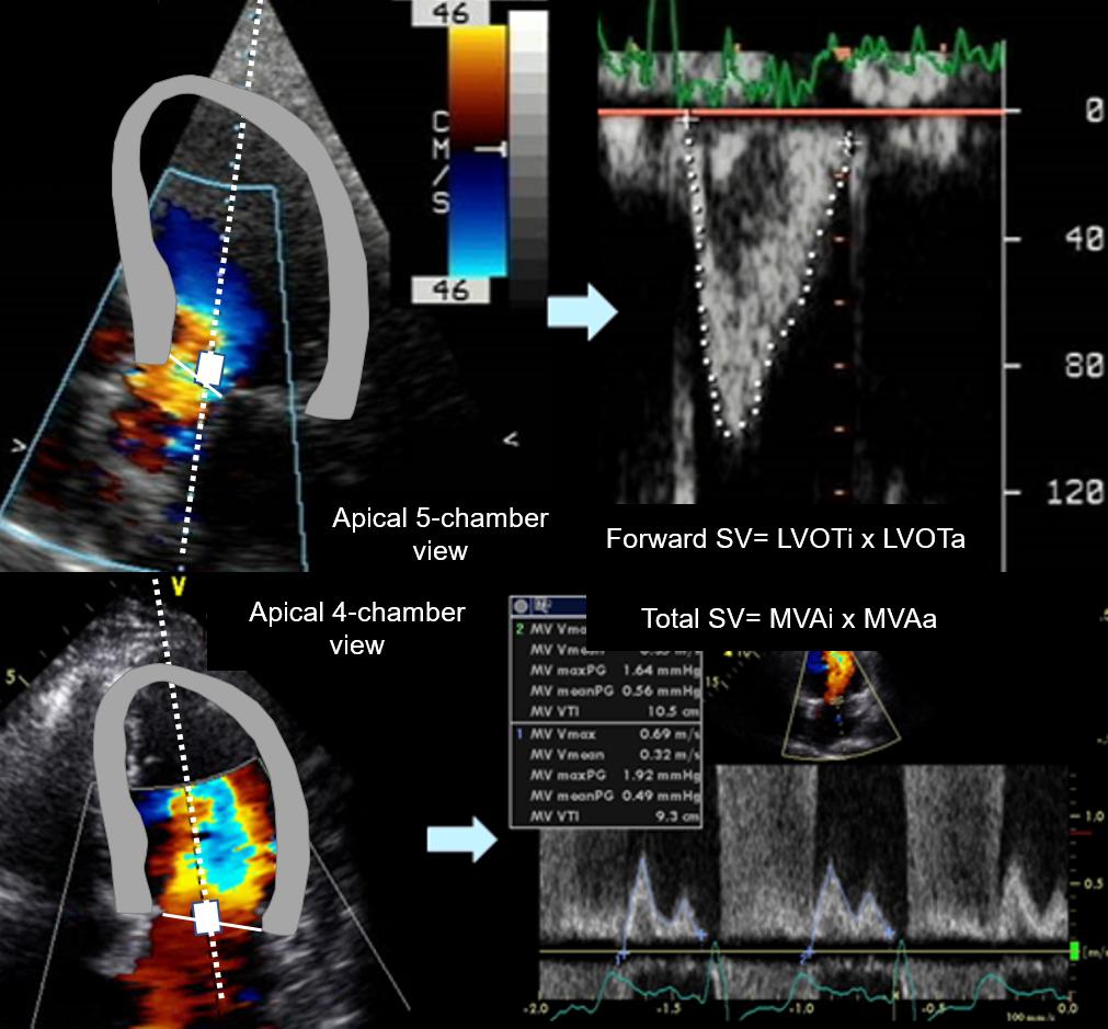
When the aortic valve does not have an associated regurgitation, the gold standard quantitative method to quantify MR is the Stroke Volume method. Mitral and aortic stroke volumes are measured with pulsed Doppler at the annular level (Figure 3), and the stroke volume of the non-regurgitant aortic valve (= forward stroke volume) is subtracted from the stroke volume of the regurgitant mitral valve (= total stroke volume = forward stroke volume + regurgitant volume), resulting in the mitral regurgitant volume. The latter can be divided by total stroke volume to obtain the regurgitant fraction.
References
Lewis JF, Kuo LC, Nelson JG, Limacher MC, and Quinones MA. Pulsed Doppler echocardiographic determination of stroke volume and cardiac output: clinical validation of two new methods using the apical window. Circulation 1984;70:425-431.
Enriquez-Sarano M, Bailey K, Seward J, Tajik A, Krohn M, Mays J. Quantitative Doppler assessment of valvular regurgitation. Circulation.1993; 87:841-848.
Figure 3. Upper panel: pulsed Doppler sampling of LVOT flow velocities in the apical 5-chamber view. Tip: note the sample volume positioned towards the mitral valve, away from the accelerated flow parallel to the base of the interventricular septum (sampling the latter location would lead to overestimation of stroke volume). Lower panel: pulsed Doppler sampling of mitral flow velocities at the annular level in the apical 4-chamber view. Tip: it is important to position the sample volume exactly in the mid annulus position in early diastole.
The imaging specialist working in the hospital Heart Team should use the Stroke Volume method whenever the regurgitant grade is not evidently severe (as in the case of a flail leaflet) since in most cases the distinction between indication/no indication for valve surgery depends on the echocardiographic distinction between severe/moderate regurgitation. It is important for the imaging specialist to be accurate and reproducible with this method, and a learning curve of a few months is necessary.
Tip: start by applying this method to normal aortic and mitral valves: the regurgitant volume should always be <10 ml. Consider that an accurate calculation of LVOT stroke volume is also necessary to calculate aortic valve area with the Continuity Equation in aortic stenosis.