How-to: Quantification of Mitral Regurgitation by the Guidelines
Quantification of mitral regurgitation with the stroke volume method
March 29, 2022
(updated April 25, 20922)
Pages 1 – 4
Topics:
- Mitral valve regurgitation
- Vena contracta
- PISA method
- Stroke volume method
- Mitral valve prolapse
- Doppler quantification
The semi-quantitative analysis of mitral regurgitation (MR) using the Vena Contracta (VC) or the PISA (Proximal Isovelocity Surface Area) method may have serious limitations.
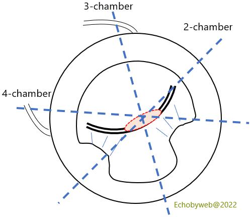
The VC (with a 6.5 mm cutoff) has been validated for a monoplane 3-chamber approach (Figure 1) or a biplane 3-chamber + 2-chamber approach, but is often used with a monoplane 4-chamber view, with a potential to overestimate functional MR because in this setting the regurgitant jet may be markedly oval (in line with the regurgitant orifice) with the major axis along the inter-commissural line (apposition line of the leaflets) (Figure 2, red oval). Further, its use is limited when the MR jet is asymmetric or spreads along the left atrial (LA) wall (Coanda effect).
The PISA method, on the other hand, is known to underestimate functional MR and its use is limited when the converging flow area impinges on the LV wall (ex: asymmetric jets arising near the mitral commissures).
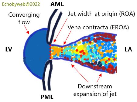
AML: anterior mitral leaflet; EROA: Effective Regurgitant Orifice Area; PML: posterior mitral leaflet; ROA: Regurgitant Orifice Area (Anatomic or Geometric Orifice Area).
References
Grayburn PA et al. Multiplane transesophageal echocardiographic assessment of mitral regurgitation by Doppler color flow mapping of the vena contracta. Am J Cardiol 1994; 74:912-917.
Roberts BJ, Grayburn PA. Color flow imaging of vena contracta in mitral regurgitation: technical considerations. J Am Soc Echocardiogr 2003;16:1002– 6.