[Page 6]

Quantitative evaluation of mitral regurgitation using the Stroke Volume method. See also our Online Calculator here.
This method is based on the measurement of:
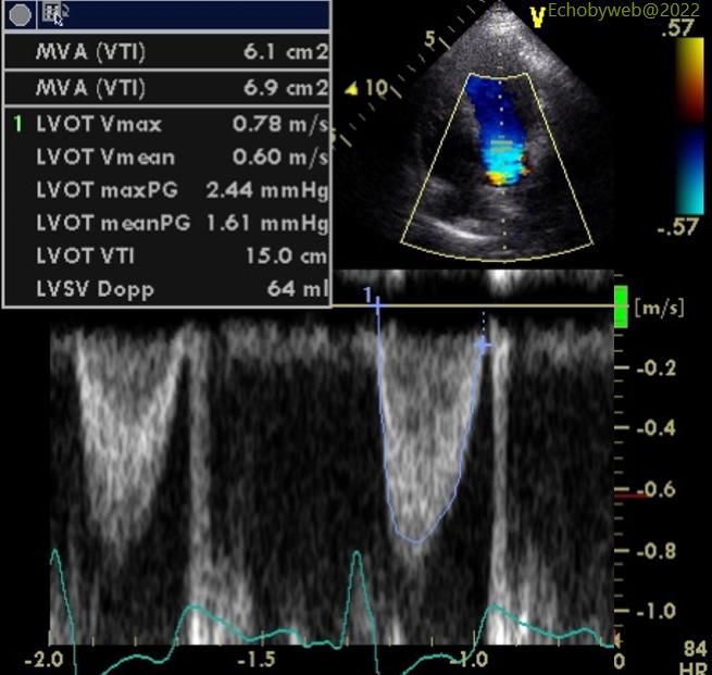
1. LV (forward) stroke volume (Figures 15-16)= LVOT area x LVOT velocity-time integral = 4.15 cm2 x 15 cm = 62.3 ml.

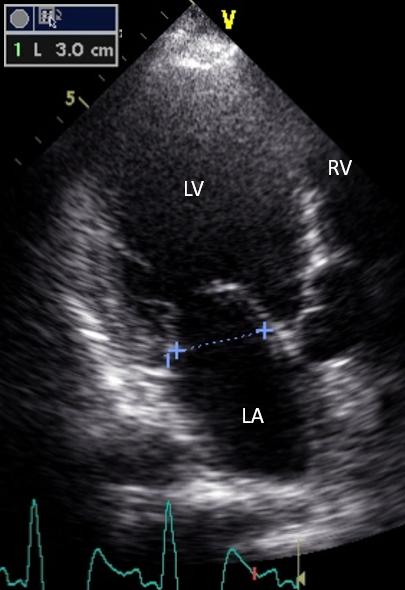
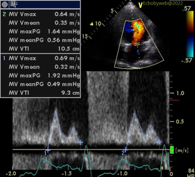
2. MItral stroke volume (Figures 17-20)= mitral annulus area x mitral annulus velocity-time integral (Figure 20) = 9.4 cm2 x 9.9 cm = 93.3 ml
3. Regurgitant Volume= mitral stroke volume – forward stroke volume = 93.3ml – 62.3 ml = 31 ml
4. Regurgitant Fraction= Regurgitant Volume / mitral stroke volume = 31 ml / 93.3 ml = 33.2 %
5. EROA= Regurgitant Volume / mitral regurgitant velocity-time integral = 31 ml / 123 cm (Figure 14) = 0.24 cm2
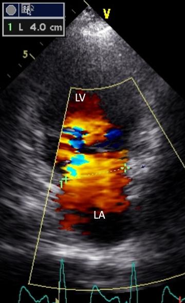
-> The mitral annulus area is calculated assuming an an ellipse geometry using the maximum (inter-c0mmissural) and minimum (antero-posterior) diameters. The former is somewhere between the 4-chambers and 2-chambers views and must be searched for. The latter is measured in the 3-chambers view. Figure 19 shows a more precise measurement of the intercommissural diameter with the help of color Doppler to define the hinge points of the mitral leaflets.
-> The mitral annulus pulsed Doppler velocity profile (Figure 20) should be searched for in the 4-chamber view by carefully positioning the sample volume at the annulus level in early diastole (when most of the diastolic flow occurs).
Since both the PISA and the Stroke Volume methods arrive at the same conclusions through independent paths, we are pretty sure about our diagnosis of moderate mitral regurgitation.