[Page 5]
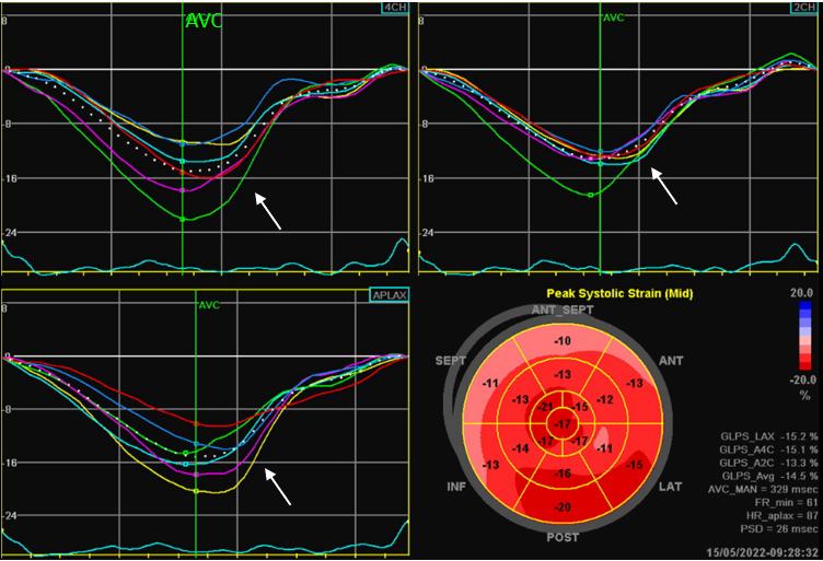
Figure 14. Left ventricular segmental longitudinal strain analysis (AFI, GE Automated Function Imaging).
The advantage of the AFI imaging software is the possibility to analyze quickly, on-board LV longitudinal segmental and global (GLS) strain. Global Longitudinal Strain is mild to moderately reduced (GLS= -14.5 %) with diffusely distributed (base to apex) lower values (compare to normal AFI below, Figure 15). LV relaxation is prolonged as shown by the reduced early diastolic segmental slopes (white arrows), but this is not different from a similarly aged normal subject with normal LV (Figure 15).
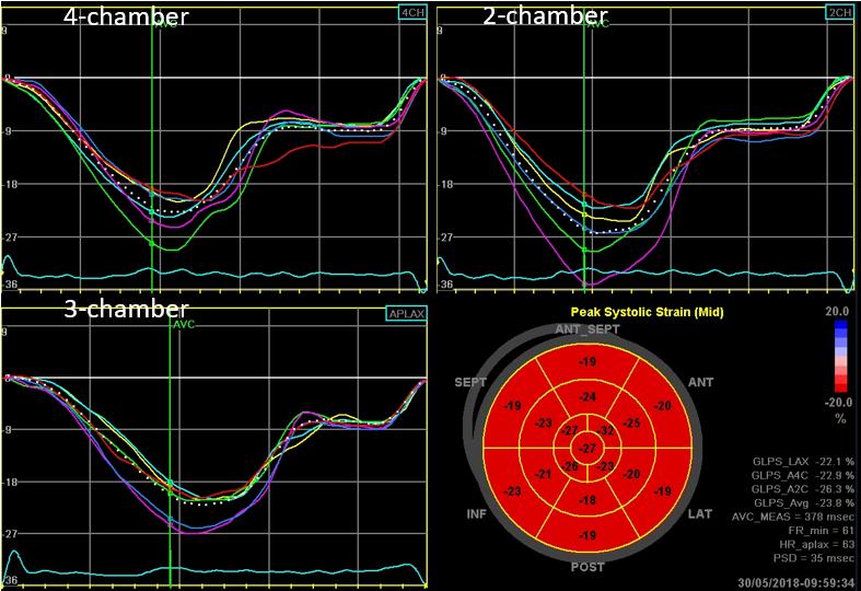
Figure 15 shows an example of normal AFI analysis of longitudinal strain in a 61 year old female normal subject (HR= 63 bpm; BSA= 1,.7 m2; BP= 120/70 mmHg). The early diastolic segmental strain profiles show a physiologic prolongation of LV relaxation as expected with ageing.
Normal LV longitudinal segmental strain values can be found in this website here.
Because of the combined dilatation of the ascending aorta (50 mm) and bicuspid aortic valve, the patient underwent replacement of the ascending aorta and aortic valve replacement with a bileaflet mechanical prosthesis. See the pre-CPB TE images here.