[Page 2]

Figure 5. 2D parasternal short axis aortic valve
Figure 6. 2D apical 5-chambers, aortic valve

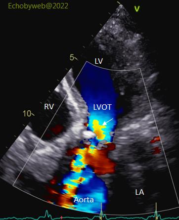
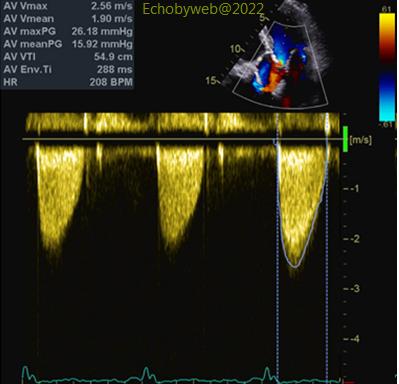
Bicuspid aortic valve with nodular calcifications (Figure 5-6), and low trans-stenotic gradient (Figure 8). The white arrow in Figure 7 shows the pre-stenotic outflow acceleration, useful to identify the stenotic orifice, so to correctly align the continuous wave Doppler to optimize sampling of the stenotic jet (Figure 8).

Figure 7. Color Doppler apical 5-chambers, aortic valve
Figure 8. Continuous wave Doppler, apical 5-chambers, aortic valve