[Page 2]
This is the transthoracic exam performed in the post-operative ICU, 1 hour after the first visualization of the dissecting hematoma in the operating room. The
hematoma has expanded and is now occupying the whole interatrial septum, protruding on both sides into the atrial cavities, as visualised in the subcostal 4-chamber view (Figures 7-8).
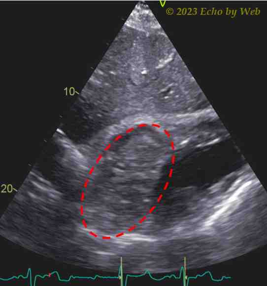
The color Doppler examination (Figure 9) does not evidence any obstruction to the venous atrial inflows or the ventricular (atrio-ventricular valves) inflows. In the apical 4-chamber view (Figure 10) the very large hematoma of the interatrial septum can be clearly seen, which also occupies most of the left atrial cavity.
The heart team decides for an observational strategy, the hemodynamics continue to be stable, and the patient is asymptomatic.
The patient is discharged from the post-operative ICU after 10 days with a good LV function; after 2 months a complete regression of the hematoma is seen on the transthoracic follow-up echocardiographic examination.