Pericardial Effusion, Tamponade and Constriction
Pericardial physiology and pathology: lesson on pericardial effusion, tamponade and constriction.
April 8th, 2022
Pages 1 – 14
Table of Contents
- Normal pericardial anatomy
- Pericardial compressive syndromes
- Pericardial adhesions & pericardial fat
- Pericardial effusion
– Estimation of severity
– Examples of effusion (mild to severe) - Pericardial tamponade
– RA wall collapse
– RV wall collapse
– Swinging heart
– Respiratory variations in flow
– Atypical post-operative presentation
– Echocardio-guided pericardiocentesis - Pericardial constriction
– Pulsed Doppler analysis
– Epidemiology
– Classification of constrictive pericarditis
– Differential diagnosis (restrictive cardiomyopathy)
– Prognosis
[See page links at bottom of page]
Normal Pericardial Anatomy
Anatomy of the pericardium.
The heart muscle is lined by the pericardium, a fibrous bag made of a white double membrane which contains a few cc of liquid (both as a “lubricant ” and as a protection from infections). The pericardium is a thin membrane of mesodermal origin that surrounds the heart, about 20 µm thick, which consists of 2 distinct layers:
– Fibrous pericardium: the outer layer, adjacent to the pleura in the mediastinum, adheres inferiorly to the diaphragm, is in continuity with the adventitia of the great vessels, and forms fibrous connections to the nearby organs (pericardial ligaments to the sternum, adhesions to mediastinal pleura, and the central tendon of the diaphragm)
– Serous pericardium: internal layer that adheres perfectly to all the flat parts and inlets of the heart. It is made of 2 leaflets:
1) Parietal. The parietal leaf consists of a layer of mesothelial cells and a fibrous collagen layer secreted by particular pericardiotic cells.
2) Visceral. The visceral leaf (epicardium) consists only of a layer of mesothelium, at the origin level of the large vessels of the vascular pedicle, which overlays all the heart.
In correspondence with the large blood vessels leaving the heart, the two parietal and visceral leafs merge.
The potential space between the parietal and visceral layers contains a thin film of fluid and is known as the pericardial cavity (20 to 50 ml of serous clear liquid: pericardial liquor)
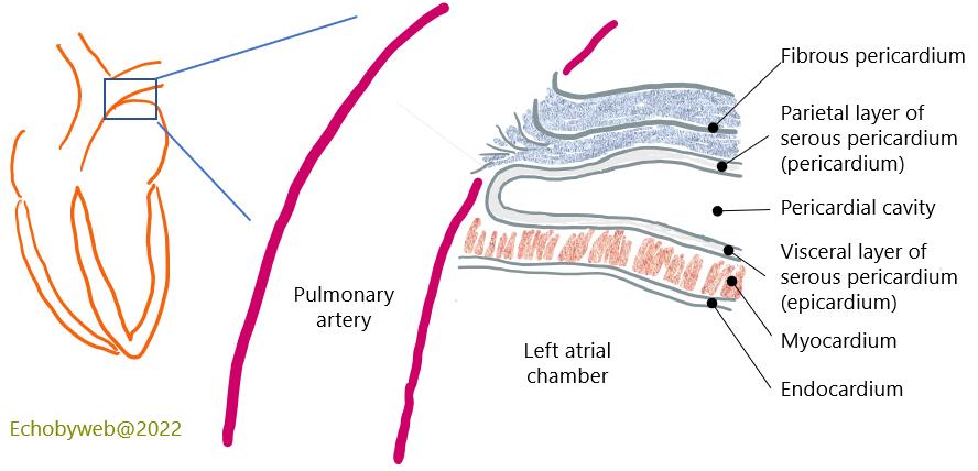
Composition of the pericardium (Figure 1).
The Transverse sinus is a space-delimited by the pericardium which is located between the aorta and pulmonary truncs (superiorly) the atria, and the superior vena cava, whereas the Oblique sinus is a space delimited by the pericardium located between the pulmonary veins.
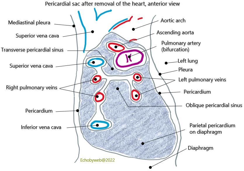
The postero-inferior pericardial sac and the pericardial sinuses: Transvere and Oblique (Figure 2).

The Transverse and Oblique pericardial sinuses as viewed from the left (Figure 3) and anterior view of the pericardial sac (Figure 4).
Functions of the pericardium.
- Primary defense mechanism against infections
- Allows the heart to adhere solidly within the anterior mediastinum
- Limits the distensibility of the heart
- The intrapericardial liquid provides lubrication and reduces friction of the muscle fibers during heart activity
- Modulates ventricular interdependence