[Page 3]
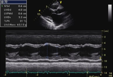
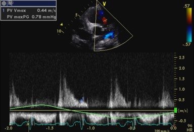
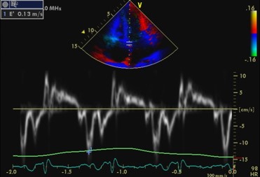
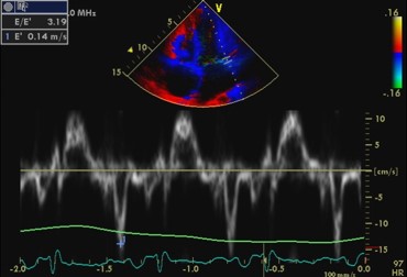
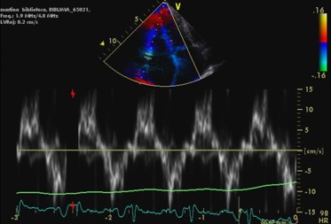
Notwithstanding the constrictive pericarditis pathophysiology, tissue Doppler imaging of the mitral annulus does not show the septal-lateral inversion of the peak velocities expected in this pathology: lateral velocities are physiologically higher than septal (Figs 14-15). The transmitral flow velocity profile is as expected in a young patient with a prevalent peak E velocity determined by normal LV relaxation, as evident from the higher septal and lateral e’ velocities (Figs 14.-15). Mean LA pressure is probably normal with E/e’ ratio= 3.2. The end-diastolic pulmonary valve gradient is low (Fig. 11). The RV tissue Doppler velocities are normal.
![M-mode echocardiogram of the left ventricle with mild posterior effusion](https://echobyweb.com/wp-content/uploads/2022/04/tt3206_ttmmlv.jpg)
![Continuous wave Doppler of pulmonary regurgitation](https://echobyweb.com/wp-content/uploads/2022/04/tt3206_ttcwpssapr.jpg)
![Pulsed Doppler mitral valve flow](https://echobyweb.com/wp-content/uploads/2022/04/tt3206_ttpdap4cmvf.jpg)
![Pulsed Doppler pulmonary venous flow](https://echobyweb.com/wp-content/uploads/2022/04/tt3206_ttpdap4cpvf.jpg)
![Tissue Doppler velocities, septal mitral annulus](https://echobyweb.com/wp-content/uploads/2022/04/tt3206_tttdilvmed.jpg)
![Tissue Doppler velocities, lateral mitral annulus](https://echobyweb.com/wp-content/uploads/2022/04/tt3206_tttdilvlat.jpg)
![Tissue Doppler velocities, lateral tricuspid annulus](https://echobyweb.com/wp-content/uploads/2022/04/tt3206_tttdirvlat-1.jpg)