[Page 3]
Outpatient transesophageal examination (Imaging is courtesy of dr. Liu Guangyu):
During the TE exam, the patient was tachycardic (> 100 bpm) and hypertensive (180/90 mmHg).
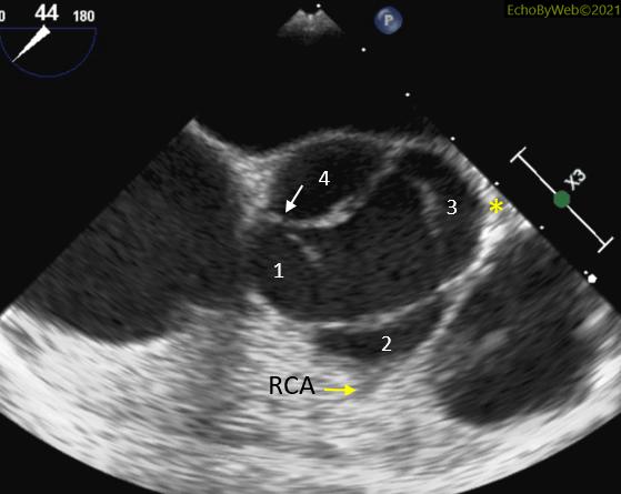
There is a quadricuspid valve with apparent incomplete diastolic apposition of the cusps (Figures 8-9). There appears to be an incomplete fusion at the base of the 2 medial cusps (towards the interatrial septum).
At color Doppler, there is apparent severe regurgitation with a wide jet extending to the apex of the LV (Figure 11). However, after shifting the aliasing velocity limit, the PISA area does not appear to be very large (i.e., not consistent with severe regurgitation) (Figure 12). The PISA calculation yielded: Regurgitant volume= 50 ml; EROA= 0.3 cm2 (consistent with moderate regurgitation).