[Page 3]
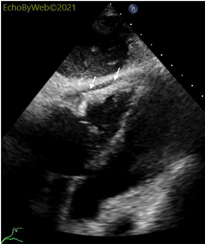
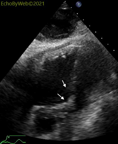
> In the subcostal 4-chamber view (Figures 8-9) it is possible to visualize in detail the anatomy and function of the pericardium lateral to the right heart chambers (white arrows):
1. no intrapericardial physiologic fluid is seen;
2. the pericardial space is occupied by fibrotic tissue;
3. there is no physiologic “sliding” of the pericardium on the epicardium during the cardiac cycle (compare to normal pericardial physiology in Figure 11).
>剑下4腔图(图8-9)可以清晰看到右心腔侧壁处心包的具体解剖结构和功能(白色箭头):
- 心包内未见生理性液体;
- 心包腔被纤维化组织占据;
- 在心动周期中,心包在心外膜上没有生理性“滑动”(与图11中的正常心包生理性相比)。