[Page 3]
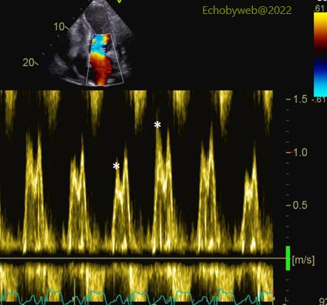

The mitral valve flow velocity profile (Figure 7) shows a marked respiratory variation of flow during respiration (asterisks: peak E wave); However, LV outflow does not decrease significantly during respiration (Figure 8). In summary, the exam shows that the moderate pericardial effusion localized lateral to the right ventricle is hemodynamically significant, but has not yet reached the point to be defined as tamponade.
二尖瓣血流速度图(图7)显示呼吸过程中明显的血流变化(星号:E波峰值);然而,呼吸过程中左室流出量并未明显减少(图8)。综上所述,检查显示局部中度心包积液,位于右室侧壁处,具有严重的血流动力学影响,但尚未达到心包填塞的定义程度。
The patient was asymptomatic at rest (in hospital post-operative period) and managed conservatively.
Figure 9 shows an almost complete regression of the effusion lateral to the right heart, after 1 week. The right atrial wall shows only active atrial contraction. All the pre-tamponade echocardiographic signs described above have regressed.
患者静息状态时无症状(术后住院期间),保守治疗。 图9显示1星期后,右心侧壁处积液几乎完全消退。右房壁只显示自主心房收缩。上述所有即将发展为心包填塞的心彩信号均已消退。