[Page 2]
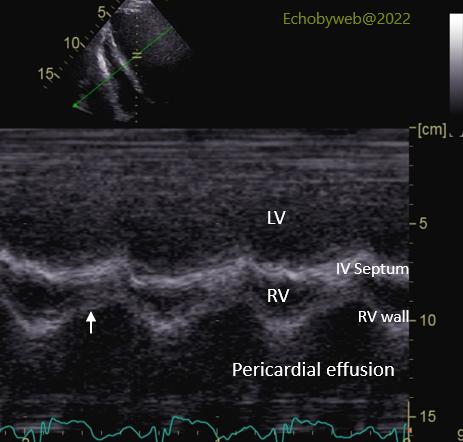


Figure 4. By using the anatomical M-mode feature (of the GE Vivid 9 machine) we align the M-mode beam perpendicular to the interventricular septum, right ventricular (RV) chamber and RV lateral wall. The white arrow points to the early to mid diastolic “collapse” of the RV wall, a clear sign of hemodynamic impairment (see here and here).
Figure 5. The same exam can be performed in the subcostal view. In addition to the diastolic collapse of the RV wall (white arrow), we can also observe the abrupt leftward shift of the interventricular septum during inspiration (dashed white arrow; respiratory tracing not recorded).
Figure 6. In the 4-chamber view, the anatomical M-mode has also been positioned perpendicular to the interatrial septum, to analyze the collapse of the right atrial wall.
图4. 应用M模解剖特征(GE Vivid 9机型),使M模线束垂直于室间隔、右室(RV)腔、以及右室侧壁。白色箭头指向舒张期初期到中期右室壁的“塌陷”,这是血流动力学受损的明确信号(见此处,及此处)。
图5. 同一个检查可以在剑下试图操作。除了右室壁舒张期塌陷以外(白色箭头),我们还能观察到吸气时室间隔突然左移(白色虚线箭头;呼吸轨迹未记录)。 图6. 在4腔图里,解剖学M模置于垂直房间隔的位置,用以分析右房壁塌陷。
The right atrial wall moves inward with a first peak at end-diastole (short white arrow), which represents the physiologic atrial contraction), whereas the second peak represents an early systolic collapse (long white arrow).
舒张末期第一个波峰处(白色短箭头),右房壁向内运动,这表示生理性心房收缩,而第二个波峰表示收缩早期塌陷(白色长箭头)。