[Page 3]
Intraoperative Images 术中图像
![Figure 10. Intraoperative anatomy pictures, before aorta clamping](https://echobyweb.com/wp-content/uploads/2021/02/te0383_or1.jpg)
![Figure 11. Intraoperative anatomy pictures, aortic clamping](https://echobyweb.com/wp-content/uploads/2021/02/te0383_or2.jpg)
![Figure 12. Intraoperative anatomy pictures, opening of aorta](https://echobyweb.com/wp-content/uploads/2021/02/te0383_or3-1024x638.jpg)
![Figure 13. Intraoperative anatomy pictures, dissection visible](https://echobyweb.com/wp-content/uploads/2021/02/te0383_or4.jpg)
![Figure 14. Intraoperative anatomy pictures, dissection visible](https://echobyweb.com/wp-content/uploads/2021/02/te0383_or5.jpg)
![Figure 15. Intraoperative anatomy pictures, dissection visible](https://echobyweb.com/wp-content/uploads/2021/02/te0383_or6.jpg)
![Figure 16. Intraoperative anatomy pictures, dissection visible](https://echobyweb.com/wp-content/uploads/2021/02/te0383_or7.jpg)
![Figure 14. Intraoperative anatomy pictures, positioning of ascending aorta graft](https://echobyweb.com/wp-content/uploads/2021/02/te0383_or8.jpg)
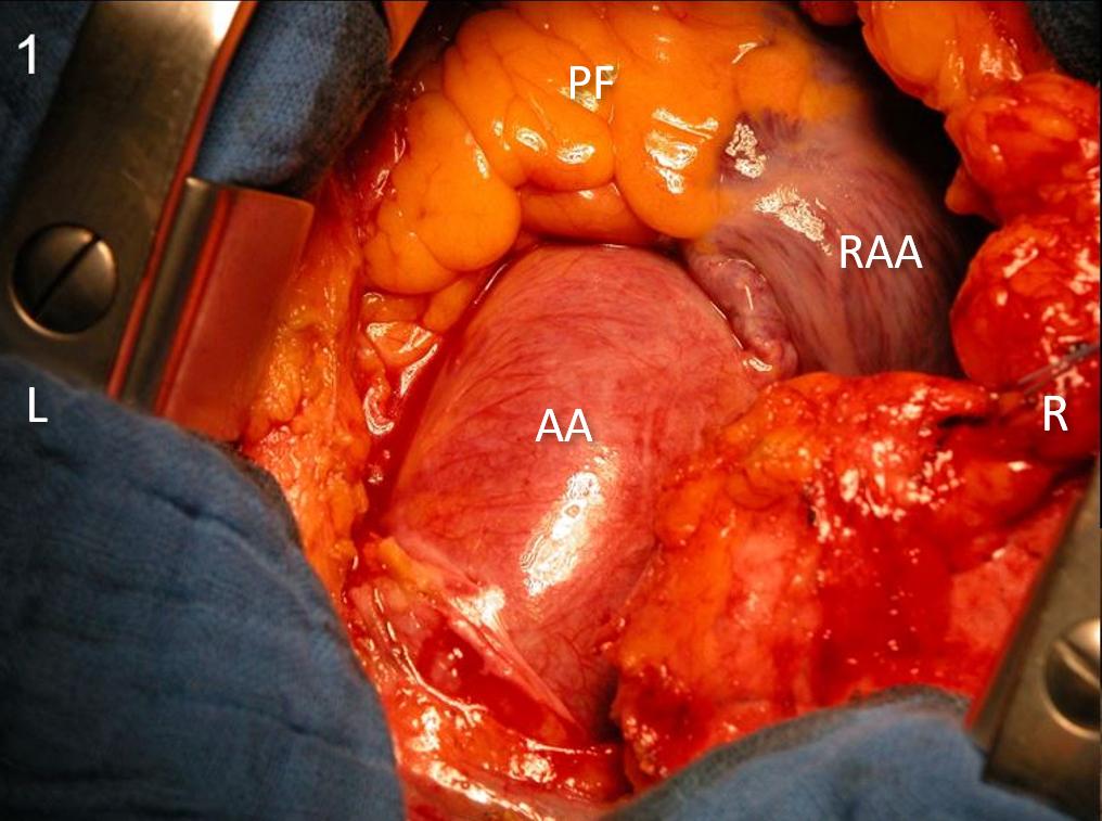
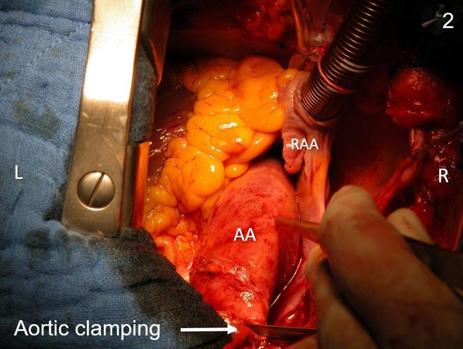
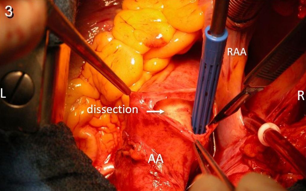
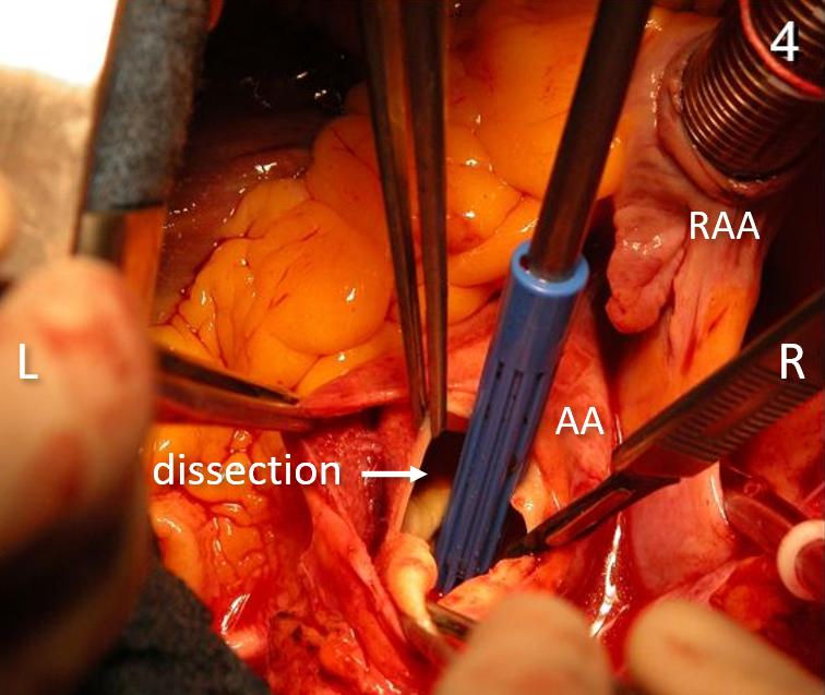
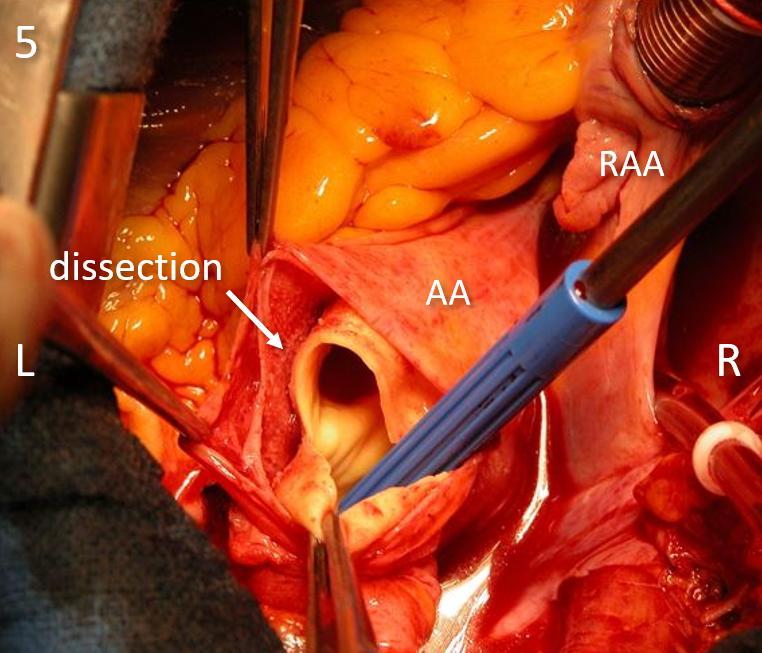
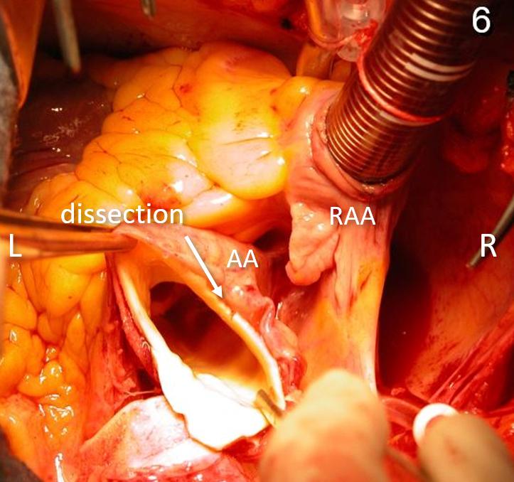
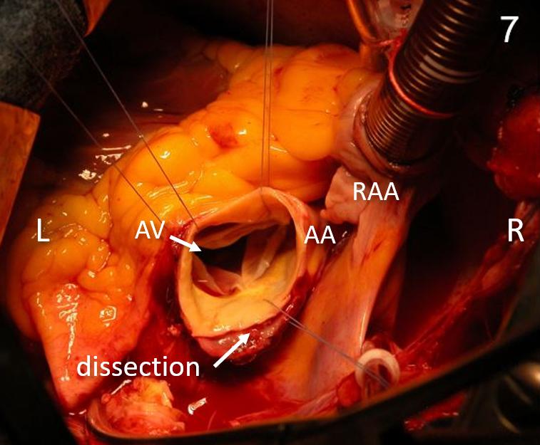
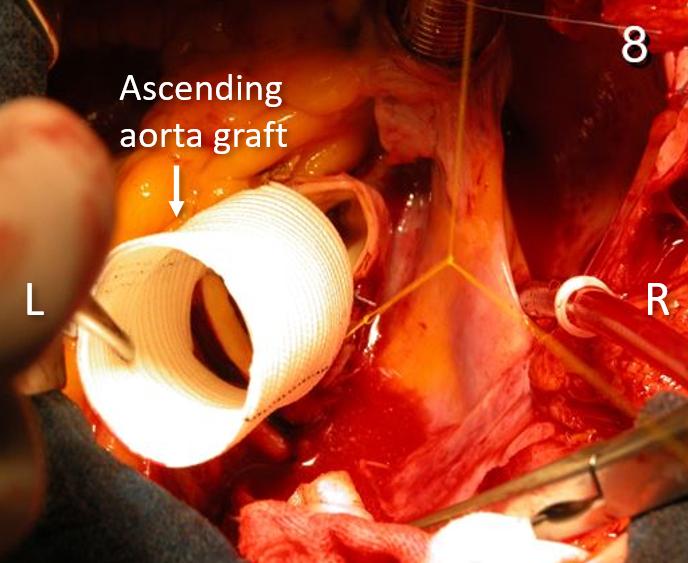
Pictures taken from the head of the patient. During clamping of aorta (Fig. 2), dissection extends to include all the aortic arch. At surgery, aortic valve was preserved; replacement of ascending aorta (Fig. 8) and arch.
AA: ascending aorta; AV: aortic valve; L: left; PF: pericardial fat; R: right; RAA: right atrial appendage.
图片从患者头部角度拍摄。主动脉阻断进行中,(图2),夹层范围遍及整个主动脉弓。手术中,主动脉瓣得以保留,置换了升主动脉(图8)及主动脉弓。
AA:升主动脉; AV:主动脉瓣; L:左; PF:心包脂肪; R:右; RAA:右心耳