{Page 2]
2D scanning of the dilated left atrium (Figure 2) showed 2 apparent thrombi inside the left atrial appendage and on the lateral atrial wall, beneath the opening of the appendage.
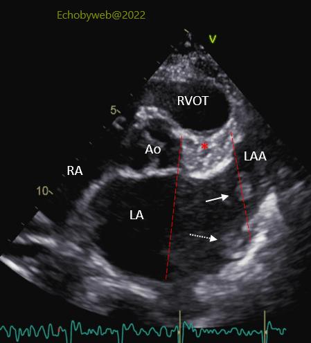
However, at the surgical inspection of the left atrial cavity during mitral valve replacement surgery, only the thrombus in the appendage was found (Figure 2, white arrow).
At a closer inspection of the 2D imaging of the dilated left atrium (Figures 2 and 3), there appeared to be an area of the lateral left atrium with ultrasound shadowing (atrial cavity section between the dashed red lines) by the tissue (red asterisk) surrounding the proximal coronary arteries, between the aortic root (Ao), right ventricular outflow tract (RVOT) and left atrial appendage (LAA). The false thrombus (Figure 2, dashed white arrow) was imaged within this area.