[Page 3]
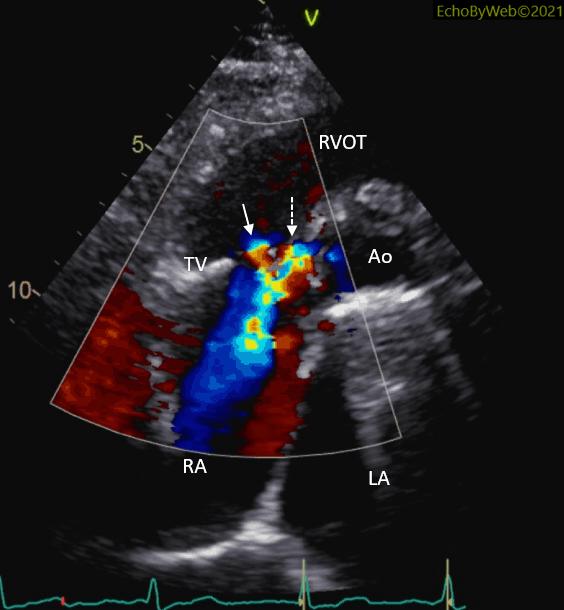
Careful angulation of the probe in color Doppler modality (in the short axis view of the tricuspid annulus and aortic root, Figure 9) shows the origin of the 2 jets (Figure 10): the first originating from the tricuspid valve (central position; white solid arrow); the second originating from the aortic root (white dashed arrow). The 2 jets after the origins tend to fuse inside the right atrial cavity.
Thus, the tricuspid regurgitation lower velocity jet points at a normal right ventricular- atrial pressure difference and normal estimated pulmonary systolic pressure, whereas the high velocity jets points at the higher aorto-atrial pressure difference (130 mmHg).
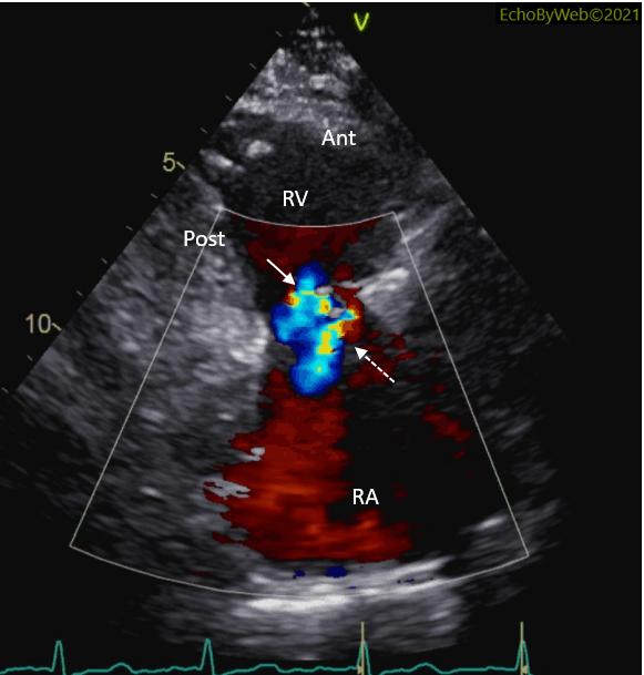
The 2 separate jets could also be visualized in the parasternal long-axis view of right ventricular inflow (Figures 11-12). White solid arrow: tricuspid regurgitation; white dashed arrow: Gerbode acquired defect.