[Page 2]
Systolic excursion of aortic root
Systolic excursion of the aortic root
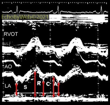
Figure 4. The systolic anterior motion of the aortic root (AO, during S) is proportional to cardiac output. The diastolic backwards motion of the aortic root is determined by the emptying dynamics of the left atrium (LA) during left ventricular filling and thus also reflects the left ventricular diastolic filling pattern.
AO: Aortic root; LA: Left atrium; RVOT: Right ventricular outflow tract; S: Systolic time interval (includes isovolumic contraction, ejection time and isovolumic relaxation). R: Early rapid left ventricular filling; C: Conduit left ventricular filling; A: Left ventricular end-diastolic filling (atrial contraction).
Calibration distance between white vertical dots = 10 mm
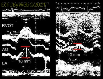
Figure 5. Left panel: systolic anterior aortic root motion in a normal subject (= 18 mm). Right panel: reduced systolic anterior aortic root motion (= 10 mm) in a patient with reduced left ventricular systolic function.
Short axis fractional shortening
Calibration distance between white vertical dots = 10 mm
M-Mode examination of the left ventricle using short axis diameters.
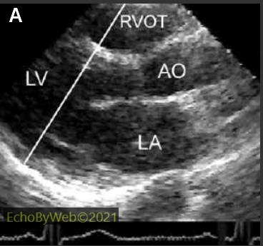
Figure 6: Correct positioning of M-mode cursor in the transthoracic parasternal long axis view to obtain the standard left ventricular M-mode tracing as shown above.
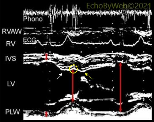
Figure 7: Measurement of LV diameters (cm).
End-diastolic at Q wave of ECG. End-systolic at either: a) end-systolic notching of IVS (yellow dashed circle); b) first wide component of II heart sound (phonocardiogram); c) maximum anterior position of the posterolateral wall.
The end-systolic notching of the IVS occurs between the maximum systolic thickening (white arrow) and the early diastolic rapid posterior movement (yellow arrow) of the anterior septum. The latter is caused by early diastolic right ventricular filling, which occurs slightly earlier than left ventricular filling, causing the posterior (passive, it is not a contraction) shift of the septum.
Fractional shortening of LV diameters
[(end-diastolic – end-systolic) / end-diastolic] x 100
LV mean velocity of circumferential fibre shortening
Vcf= (end-diastolic – end-systolic) / (ejection time x end-diastolic) (circ/sec)
IVS: interventricular septum; LV: left ventricular cavity; Phono: phonocardiographic tracing; PLW: postero-lateral wall; RV: right ventricular cavity; RVAW: RV anterior wall; RVOT: RV outflow tract.
Left ventricular wall thickening
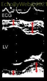
Figure 8. Measurement of end-diastolic (red arrows synchronous with Q wave of ECG) and end-systolic (at maximum systolic wall thickness) left ventricular wall thickness.
Wall thickening (%): [(end-systolic thickness – end-diastolic thickness) / end-diastolic thickness] x 100
Measurement units: cm. Normal values:
Interventricular septum= 34 – 50 %
Posterior wall= 43 – 61 %