[Page 5]
Relevance of the ECG trace.
- Events related to presence of P wave (arrhythmias; AV block; PM)
- Timing of morphological or flow-related events:
– Location of end-systolic LV volume in severe dyssynchrony
– To distinguish pulmonary venous flow waves
– To differentiate systolic from diastolic flow events when sampling shunts in congenital heart diseases - Possibility to obtain simultaneous respiratory tracing to evaluate influence of respiration on morphological and flow events (ex: constrictive pericarditis)
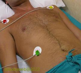
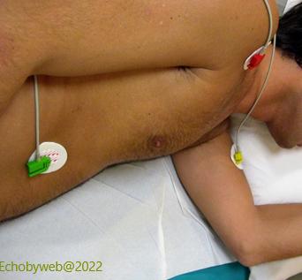
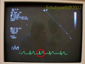
Figure 11 and 12. Optimal position of ECG electrodes in the flat and left recumbent positions. The standard position of the electrodes allows an optimal visualization of the ECG waves ( P, QRS, T) (Figure 13).
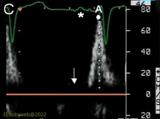
Figure 15. The presence of the ECG P wave allows us to identify the pulsed Doppler mitral flow wave as a end-diastolic “A wave”. In the absence of the ECG, the wave can be easily mistaken as an early diastolic “E” wave.
Figure 14. The availability of an ECG tracing allows to identify with greater confidence LV end-diastole to perform LV planimetry and calculate LV end-diastolic volume.
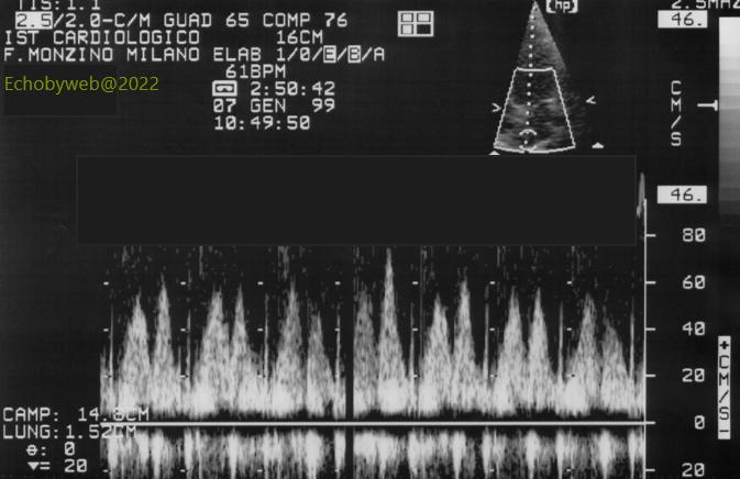
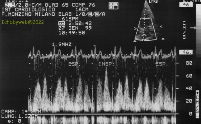
The ECG tracing allows an easier identification of the systolic and early diastolic pulmonary venous flow waves. Compare the tracings without ECG (Figure 16) and with the ECG tracing (Figure 17).
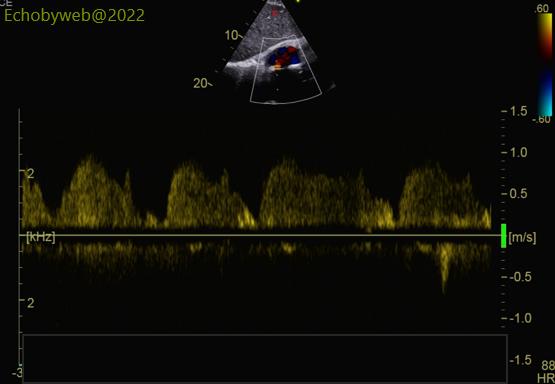
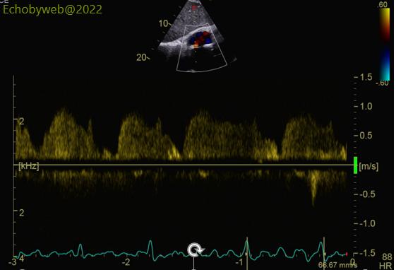
The presence of a good quality ECG tracing is essential in the case of Congenital Heart Disease. In this example, the ECG helps in identifying the different waves of a small ASD (atrial septal defect) with mild left to right shunt.