[Page 8]
RV wall diastolic collapse.
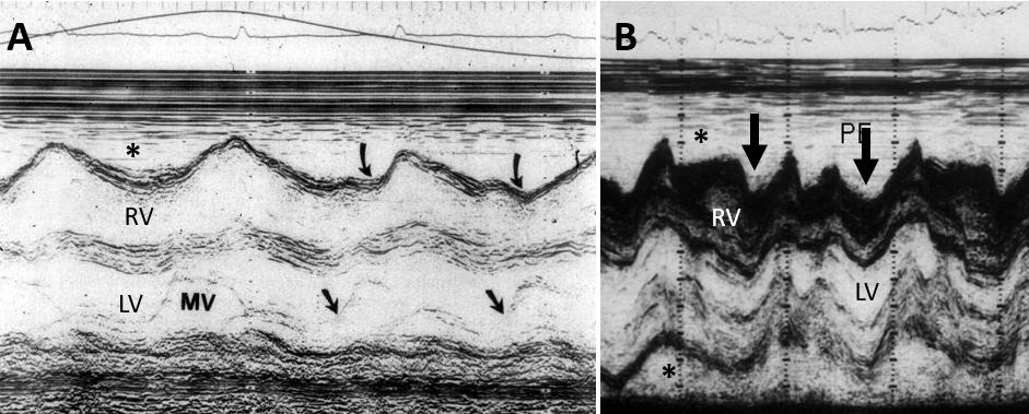
A. Partial early diastolic collapse (curved black arrow; black arrow marks beginning of diastole)
B. complete diastolic collapse of the RV free wall (thick black arrows). Asterisk: effusion.
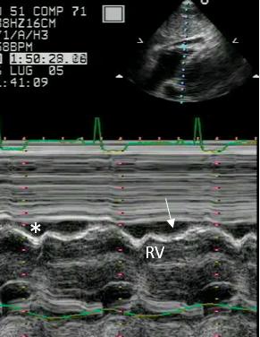
Figures 50-51. Prolonged diastolic collapse of the RV lateral wall (white arrow). Asterisk: pericardial effusion.
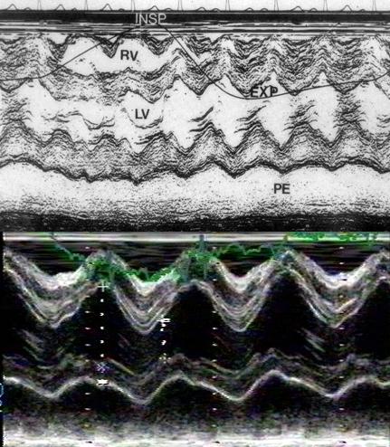
Excessive movement of the heart within the pericardial sac (the swinging heart).