2D Aortic Arch measures
2D measurement of the aortic arch from the suprasternal window
January 23, 2021
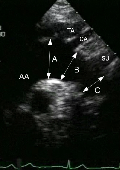
Suprasternal (jugular) 2D longitudinal view of the aortic arch.
Measures: A. proximal arch; B. mid-arch; C. distal arch. AA: ascending aorta; TA: anonymous art. (brachiocephalic); CA: left carotid; SU: left subclavian art.
Measures should be performed “inner edge” to “inner edge”. I prefer to measure the arch at the “exit” point (C, the boundary between the aortic arch and the descending thoracic aorta), which is also the (normal) smaller diameter, immediately distal to the left subclavian artery. When in doubt about the anatomy (poor image resolution), adding color Doppler may help identify the lumen of the aortic arch, and the correct diameter (gain should be kept low to avoid shadowing of the anatomy of the walls by “overflowing” color signal).
胸骨上(颈静脉)主动脉弓纵向切面2维视图
测量:A.主动脉弓近端;B. 主动脉弓中段;C. 主动脉弓远端AA:升主动脉;TA:无名动脉(头臂动脉);CA:左颈动脉;SU:左锁骨下动脉。
测量应采用“内缘”到“内缘”的操作方式。我比较倾向于在“出口”点(C)处测量主动脉弓,这也是(正常)较小的直径,紧靠左锁骨下动脉远端。当对解剖结构有疑问时(图像分辨率差),添加彩色多普勒可能有助于确认主动脉弓的管腔和其正确的直径(保持低增益值,避免管壁解剖结构由于彩色信号“过溢”产生阴影)。